Diaporthe fusiformis Jayaward & Manawas, sp. nov. 2021
MycoBank Index fungorum: IF557998; Facesoffungi number: FoF09385
Holotype: JZBH3340154.
Morphological description
Sexual morph:
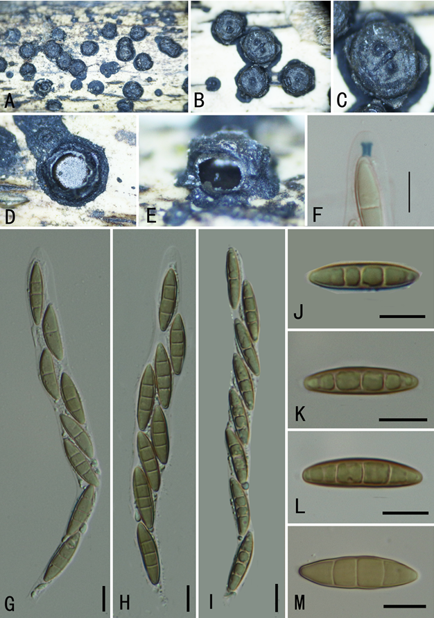
Asexual morphs: Pycnidia on PDA superficial, scattered, black, globose, solitary and clustered. Conidiophores reduced to conidiogenous cells. Conidiogenous cells hyaline, smooth–walled clustered. Alpha conidia 8–5 × 2–3 μm (x̅= 7 × 2 μm, n = 40), eguttulate, hyaline, fusiform, both ends angular. Beta conidia 23–32 ×1.2–1.6 μm (x̅= 27× 1.5 μm, n = 40), aseptate, hyaline, hamate, filiform, tapering towards both ends. Gamma conidia not observed.
Cultures: Colonies on PDA reach 90 mm diam. after five days at 25°C,producing abundant white aerial mycelia and reverse fuscous white becoming gray.
Habitat: on dead Camellia sinensis leaves.
Distribution: CHINA, Fujian Province, Zhangzhou County.
GenBank Accession: ITS:MW010218;tub2:MW056014;tef1:MW205234;CAL: MW205218.
Notes: In the phylogenetic analysis four isolates obtained in this study formed a well–supported clade with 100% ML and 0.98 PP values. In the recombination analysis, PHI test indicated that these isolates belong to a species separate from all other species in Diaporthe (Fig. 9). Diaporthe fusiformis is phylogenetically close to Diaporthe eucalyptorum (CBS132525) and Diaporthe fujianensis (This study). Diaporthe eucalyptorum (CBS132525) has larger conidia and Diaporthe fujianensis has smaller conidia (4–6 × 2–3 μm) than Diaporthe fusiformis (8–5 × 2–3 μm). In addition, the conidia of Diaporthe fusiformis are fusiform whereas Diaporthe eucalyptorum has biguttulate fusoid conidia and Diaporthe fujianensis has oval to ellipsoidal conidia. In comparison with Diaporthe lithocarpus; Diaporthe fujianensis has smaller conidia (4–6 × 2–3 μm) than Diaporthe lithocarpus (6–8 × 2–3 μm). Diaporthe lithocarpus develop both alpha conidia and beta conidia whereas Diaporthe fujianensis is prominent with alpha conidia. Based on morphological and phylogenetic characters we identified this taxon as a novel species.
Reference: Manawasinghe IS, Jayawardena RS, Li HL, Zhou YY, Zhang W, Phillips AJL, Wanasinghe DN, Dissanayake AJ, Li XH, Li YH, Hyde KD, Yan JY 2021 – Microfungi associated with Camellia sinensis: A case study of leaf and shoot necrosis on Tea in Fujian, China. Mycosphere 12(1), 430–518, Doi 10.5943/mycosphere/12/1/6
Diaporthe fusiformis (JZBH3340154 Holotype). a–c Pycnidia on PDA. d Conidiogenus cells. e–g Alpha conidia. h Beta conidia. i Upper view of colony on PDA after five days. j Reverse view of colony on PDA after five days. Scale bars: b–c = 100 µm, d = 20 µm, e–h = 10 µm.