Diaporthe fujianensis Jayaward & Manawas, sp. nov. 2021
MycoBank Index Fungorum: IF557997; Facesoffungi number: FoF09384
Holotype: JZBH3340150.
Morphological description
Sexual morph:
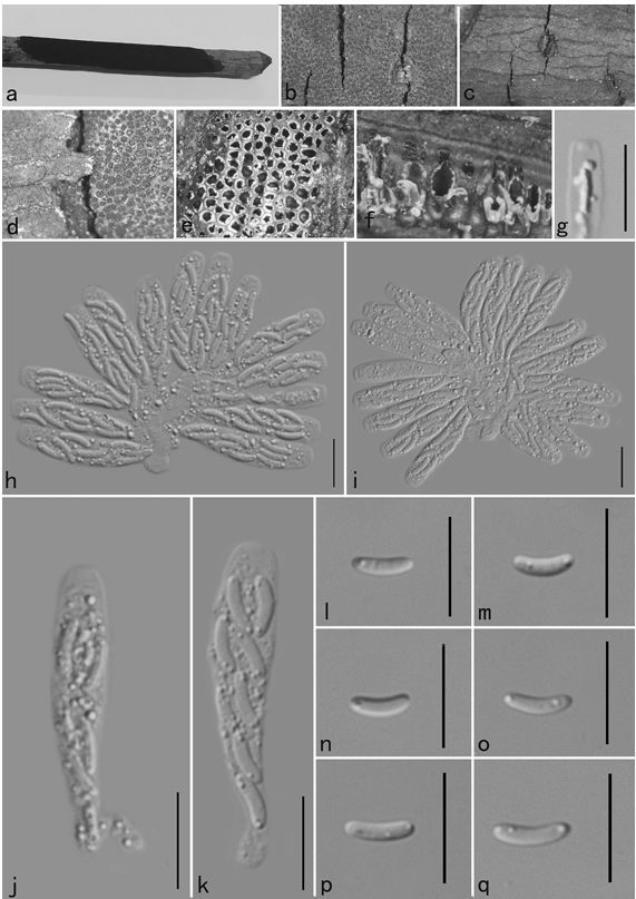
Asexual morphs: Pycnidia on PDA superficial, scattered, black, globose, solitary in most. Conidiophores not observed. Conidiogenous cells terminal, hyaline and smooth. Alpha conidia 4–6 × 2–3 μm (x̅= 5 × 2.5 μm n = 40), biguttulate, hyaline, oval and or ellipsoidal, both ends obtuse. Beta conidia and gamma conidia were not observed.
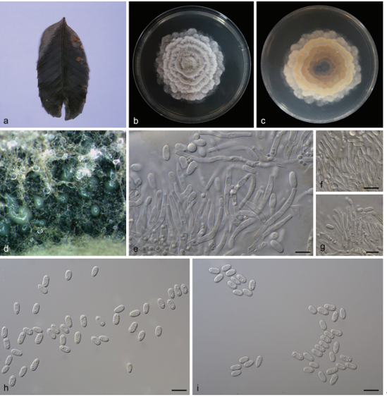
Cultures: Colonies on PDA reach 90 mm diam. after five days at 25°C, producing abundant white aerial mycelia and reverse fuscous white.
Habitat: on dead Camellia sinensis shoots.
Distribution: CHINA, Fujian Province, Zhangzhou County.
GenBank Accession: ITS: MW010212;tub2: MW056008;tef1: MW205231;CAL: MW205212.
Notes: In the phylogenetic analysis four isolates obtained in this study clustered in a well–supported clade with 100% ML and 84% MP bootstrap values and 0.98 BYPP. In the recombination analysis, PHI test indicated that the current isolates belong to a species separated from all other Diaporthe species included in the phylogenetic tree. Diaporthe fujianensis resides in a sister clade to Diaporthe eucalyptorum. Morphologically the alpha conidia produced by this species are smaller than those in Diaporthe eucalyptorum (6 × 2.5 µm). A pairwise nucleotide comparison between Diaporthe eucalyptorum ex type strain (CBS 132525) and Diaporthe fujianensis ex type strain (JZBH320150) in ITS region showed 1.75% base pair differences along 519 bp. Based on the molecular evidences we consider that these isolates belong to a novel species.
Reference: Manawasinghe IS, Jayawardena RS, Li HL, Zhou YY, Zhang W, Phillips AJL, Wanasinghe DN, Dissanayake AJ, Li XH, Li YH, Hyde KD, Yan JY 2021 – Microfungi associated with Camellia sinensis: A case study of leaf and shoot necrosis on Tea in Fujian, China. Mycosphere 12(1), 430–518, Doi 10.5943/mycosphere/12/1/6
Diaporthe fujianensis (JZBH3340150 holotype) a Diseased shoot. b Upper view on of colony PDA after five days. c Reverse view of colony on PDA after five days. d–f alpha conidia. Scale bars: d–f = 10 µm.