Diaporthe sinensis Jayaward & Manawas, sp. nov. 2021
MycoBank Index Fungorum: IF557999; Facesoffungi number: FoF09387
Holotype: JZBH320167.
Morphological description
Sexual morph:
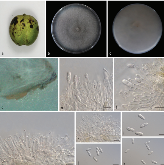
Asexual morphs: Pycnidia on PDA 360–900 μm (x̄ = 500 µm, n = 20) in diam., superficial, scattered, dark brown to black, globose, solitary in most. Conidiophores reduced to Conidiogenous cells. Conidiogenous cells hyaline, simple, smooth terminal. Alpha conidia 7–4 ×2–3 μm (x̅= 5 × 3 μm, n = 40) hyaline, oval, both ends obtuse. Beta conidia and gamma conidia not observed.
Cultures: Colonies on PDA reach 90 mm diam., after five days at 25°C, producing abundant white aerial mycelia and reverse fuscous white.
Habitat: on dead Camellia sinensis leaves.
Distribution: CHINA, Fujian Province, Zhangzhou County.
GenBank Accession: ITS: MW010224;tub2: MW056016;tef1: MW205235;CAL: MW205220.
Notes: In the phylogenetic analysis, four isolates obtained in this study formed a well–supported clade with 70% ML and 68% MP bootstrap values and 0.97 BYPP. These taxa show particular neighbour relation to Diaporthe amygdali (CBS 126679). Compared to the sister species, Diaporthe sinensis develops oval and shorter alpha conidia whereas conidia of Diaporthe amygdali are fusiform, and biguttulate (Gomes et al. 2013). A comparison of the ITS (497bp), tef1 (492bp), and Cal (300bp) between our species (JZBH3340167) and closely associated Diaporthe amygdali (CBS 126679) revealed 2%, 2.4% and 14% base pair differences respectively. Therefore, based on both morphological and phylogenetic evidence we identified these isolates as a novel Diaporthe species associated with tea.
Reference: Manawasinghe IS, Jayawardena RS, Li HL, Zhou YY, Zhang W, Phillips AJL, Wanasinghe DN, Dissanayake AJ, Li XH, Li YH, Hyde KD, Yan JY 2021 – Microfungi associated with Camellia sinensis: A case study of leaf and shoot necrosis on Tea in Fujian, China. Mycosphere 12(1), 430–518, Doi 10.5943/mycosphere/12/1/6
Diaporthe sinensis (JZBH3340167 Holotype) a Diseased leaf. b Upper view of mycelium on PDA five days. c Reverse view of mycelium on PDA five days. d Pycnidia on PDA. e–f Alpha conidia. Scale bars: e = 10 µm, f = 5 µm.