Sanguinoderma perplexum (Corner) Y.F. Sun & B.K. Cui, comb. nov. 2020
MycoBank MB828445
Holotype: China, Hainan Province, Changjiang County, 2009, B.K. Cui, Cui 6496 (BJFC); on angiosperm stump, 9 May 2009, Y.C. Dai, Dai 10811 (BJFC); ibid., 10 May 2009, B.K. Cui, Cui 6554 (BJFC).
Morphological description
Basidiomata annual, almost sessile or with a very short and thick stipe, hard corky to woody hard. Pileus single, suborbicular to flabelliform, applanate, up to 11.5 cm long, 8 cm wide and 3 cm thick. Pileal surface reddish brown to ferruginous, dull, glabrous, with concentric furrows and radial wrinkles; margin obtuse, entire, slightly wavy when dry. Pore surface pale white to cream when fresh, colour changing to blood red when bruised, then quickly darkening; pores circular to angular, 5–6 per mm; dissepiments medially thick, entire. Context yellowish brown to cinnamon, with a bunch of melanoid lines, corky, up to 2.4 cm thick. Tubes concolorous with pore surface, hard corky, up to 6 mm long. Stipe concolorous with pileal surface, lateralattached, up to 2 cm long and 3 cm diam. Hyphal system trimitic; generative hyphae with clamp connections, all hyphae IKI–, CB+; tissues darkening in KOH. Generative hyphae in context colourless, thin-walled, 3–5 μm diam; skeletal hyphae in context pale yellow to pale brown, thick-walled with a wide to narrow lumen or subsolid, arboriform branched and flexuous, 3–7 μm diam; binding hyphae in context colourless, subsolid, branched and flexuous, 1–3 μm diam. Generative hyphae in tubes colourless, thin-walled, 2–4 μm diam; skeletal hyphae in tubes pale yellow to yellowish brown, thick-walled with a wide to narrow lumen or subsolid, arboriform branched and flexuous, 3–6 μm diam; binding hyphae in tubes colourless, subsolid, branched and flexuous, 1–3 μm diam. Pileal cover composed of clamped generative hyphae, thin- to thick-walled, apical cells gelatinized, flexuous, yellowish brown, about 30–50 × 4–8 μm, forming an irregular palisade. Cystidia absent; cystidioles clavate, colourless, thin-walled, 27–30 × 2–3 μm. Basidia barrel-shaped to clavate, colourless, thin-walled, 30–44 × 22–28 μm; basidioles in shape similar to basidia, colourless, thin-walled, 19–30 × 5–11 μm. Basidiospores subglobose to broadly ellipsoid, pale yellow, IKI–, CB+, with double and medially thick walls, exospore wall smooth, endospore wall with conspicuous spinules, (11.1–)11.5–14(–14.8) × (9–)10–12(–13.9) μm, L = 12.75 μm, W = 10.84 μm, Q = 1.16–1.19 (n = 60/2). Under SEM, exospore wall densely vermiculate to semi-reticulate, endospore wall with long and thick columnar spinules loosely arranged.
Habitat: on angiosperm stump
Distribution: China. Malaysia.
GenBank Accession: ITS KJ531650; nLSU KJ531650; RPB2 MK121538a; TEF MK121583a
Notes: Amauroderma perplexum was described from Malaysia (Corner 1983). It can be mainly characterized by sessile to substipitate and woody hard basidiomata, clavate cystidioles and a vermiculate to semi-reticulate exospore wall with thick columnar endospore spinules (Fig. 8k–l). In addition, A. perplexum presents a pore surface that changes to blood red when bruised and is shown to be a distinct lineage in Sanguinoderma with high phylogenetic support (100 % ML, 1.00 BPP). Therefore, we transferred A. perplexum to Sanguinoderma.
Sanguinoderma perplexum is similar to S. laceratum by sharing broadly ellipsoid basidiospores with slightly thick walls, but S. laceratum differs from S. perplexum by its thin and lacerate dissepiments and semi-reticulate exospore wall with slightly thin and coniform endospore spinules (Fig. 8g–h).
Reference: Y.-F. Sun1,2, D.H. Costa-Rezende3, J.-H. Xing1 et al.
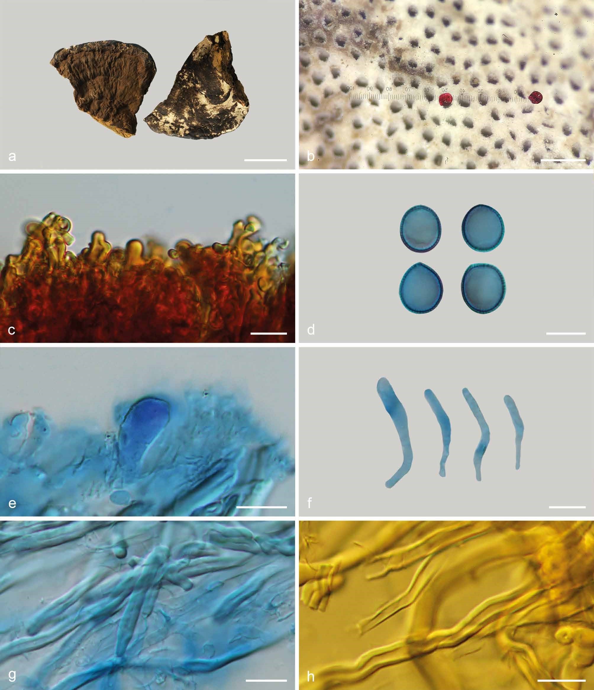
Basidiomata and microscopic structures of Sanguinoderma perplexum (Dai 10811). a. Basidiomata; b. pores; c. apical cells from pileal cover; d. basidiospores; e. basidioles; f. cystidioles; g. generative and skeletal hyphae from tubes; h. skeletal hyphae from context. — Scale bars: a = 3 cm; b = 0.5 mm; c–h = 10 µm.