Sanguinoderma microporum Y.F. Sun & B.K. Cui, sp. nov. 2020
MycoBank MB828437
Holotype: China, Hainan Province, Qiongzhong County, Limushan National Forest Park, on ground of angiosperm forest, 16 June 2016, B.K. Cui, Cui 13851 (BJFC).
Morphological description
Basidiomata annual, centrally to laterally stipitate, woody hard. Pileus single, suborbicular to flabelliform, up to 10.5 cm diam and 8 mm thick. Pileal surface pale yellowish brown to greyish brown, dull, glabrous, with concentric dark zones or furrows and radial wrinkles; margin acute to obtuse, entire, wavy and incurved when dry. Pore surface yellowish brown to dark brown when fresh, colour changing to blood red when bruised and becoming greyish brown when dry; pores circular, 6–7 per mm; dissepiments extremely thick (about 0.12–0.16 mm thick), entire. Context straw colour, with dark melanoid lines, corky, up to 4 mm thick. Tubes pale brown, up to 4 mm long. Stipe concolorous with pileal surface, cylindrical and hollow, up to 10.5 cm long and 9 mm diam. Hyphal system trimitic; generative hyphae with clamp connections, all hyphae IKI–, CB+; tissues darkening in KOH. Generative hyphae in context colourless, thin-walled, 3–4 μm diam; skeletal hyphae in context pale yellow, thick-walled with a wide to narrow lumen or subsolid, arboriform branched and flexuous, 3–8 μm diam; binding hyphae in context colourless, subsolid, branched and flexuous, up to 2 μm diam. Generative hyphae in tubes colourless, thin-walled, 2–3 μm diam; skeletal hyphae in tubes pale yellow, thick-walled with a wide to narrow lumen or subsolid, arboriform branched and flexuous, 3–6 μm diam; binding hyphae in tubes colourless, subsolid, branched and flexuous, 1–2 μm diam. Pileal cover composed of clamped generative hyphae, thin- to thick-walled, apical cells clavate, faintly inflated and flexuous, pale yellow to dark brown, about 25–40 × 6–13 μm, forming an irregular palisade. Cystidia or cystidioles absent. Basidia barrel-shaped to clavate, colourless, thin-walled, 18–22 × 13–18 μm; basidioles in shape similar to basidia, colourless, thin-walled, 16–20 × 10–15 μm. Basidiospores broadly ellipsoid, pale yellow, IKI–, CB+, with double and slightly thick walls, exospore wall smooth, endospore wall with conspicuous spinules, (10.7–)11–12(–12.3) × (8.5–)8.7–9.8(–10) μm, L = 11.52 μm, W = 9.18 μm, Q = 1.23–1.28 (n = 60/2). Under SEM, exospore wall semi-reticulate, endospore wall with long and slightly thin coniform spinules tightly arranged.
Habitat: on ground of angiosperm forest
Distribution: Hainan Province, China.
GenBank Accession: ITS MK119854a; nLSU MK119933a; RPB2 MK121512a; TEF MK121602a
Notes: Sanguinoderma microporum is a distinct species on account of its distinctly small pores with extremely thick dissepiments. It has broadly ellipsoid basidiospores with slightly thick walls which are similar to S. laceratum. But S. laceratum differs from S. microporum by its superposed pileus and lacerate pores with thin dissepiments. In the phylogenetic analyses, S. microporum was shown to be a distinct well-supported lineage in Sanguinoderma (Fig. 1, 2).
Reference: Y.-F. Sun1,2, D.H. Costa-Rezende3, J.-H. Xing1 et al.
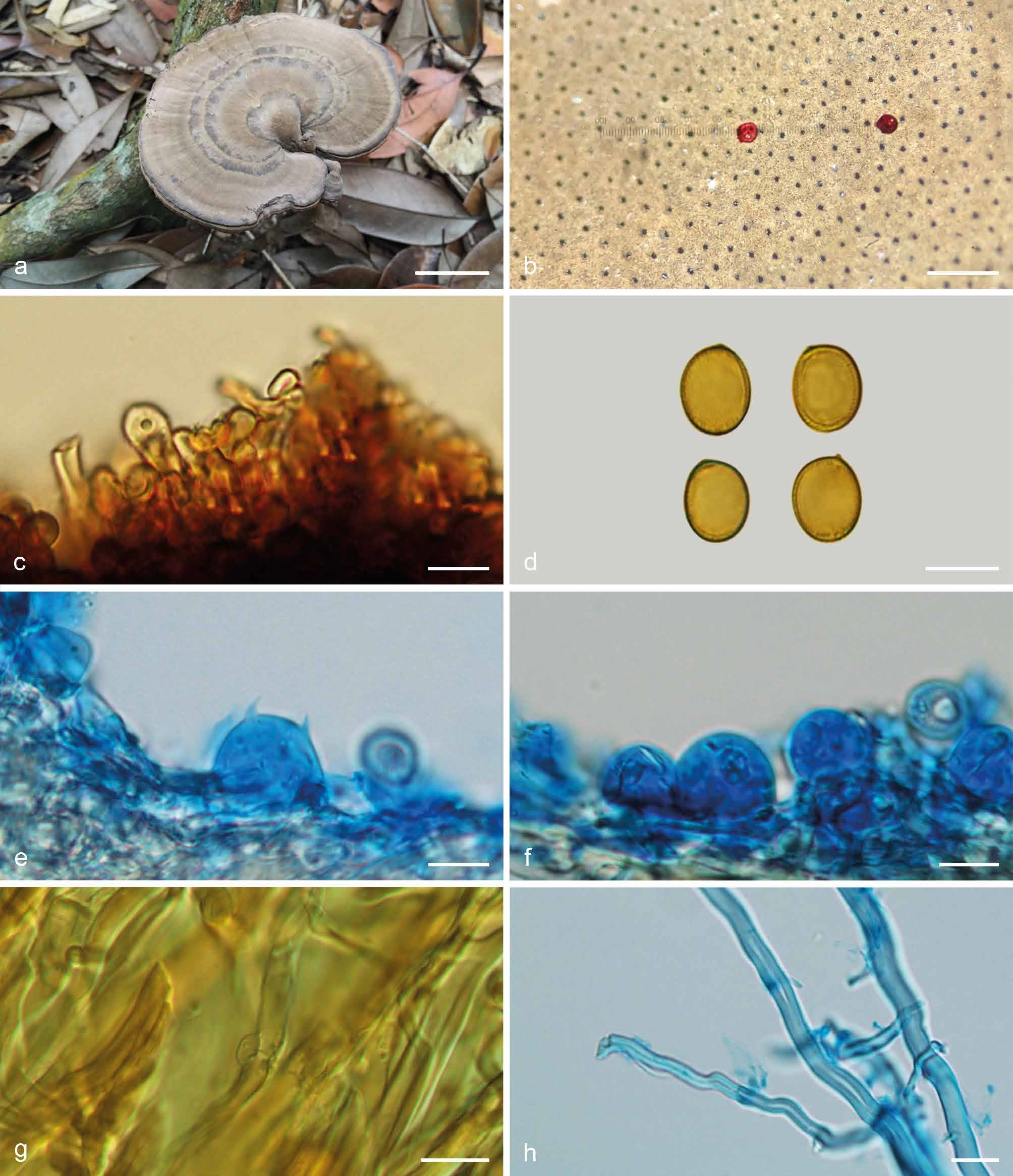
Basidiomata and microscopic structures of Sanguinoderma microporum (Cui 13851). a. Basidiomata; b. pores; c. apical cells from pileal cover; d. basidiospores; e. basidia; f. basidioles; g. generative hyphae from tubes; h. skeletal hyphae from context. — Scale bars: a = 3 cm; b = 0.5 mm; c–h = 10 µm.