Aspergillus limoniformis Z.F. Zhang & L. Cai, sp. nov. 2020
Index Fungorum number: 556394; Facesoffungi number: FoF 08427
Holotype: CHINA, Yunnan, Mengzi, Mingjiu old Cave, N 23.487°, E 103.619°, on bat guano, May 2016, Z.F. Zhang, HMAS 248014 (holotype designated here), ex-type living culture CGMCC3.19323 = LC126098; ibid., LC12610.
Morphological description
Hyphae hyaline, septate, smooth, branched, 1.0–2.5 μm wide. Asexual morph Conidiogenous cells simple phialides arising laterally on vegetative hyphae. Phialides cylindrical, ampulliform, or tapering with enlarged base, smooth, hyaline, variable in length, 4.0–10.0 µm long, 1.5–5.0 µm diam. at base, tapering to 1.0–2.0 µm diam. at apex. Conidia formed in long chains, limoniform or subglobose, obviously apiculate, thick-walled, rough initially, then becoming smooth with age, hyaline, 3.0–4.5 × 2.5–4.0 µm ( x̄ ± SD = 3.7 ± 0.33 × 3.3 ± 0.25 µm, n = 60). Sexual morph not observed. Culture characteristics—Colonies on PDA attaining 25–31 mm diam. after 4 weeks, flat, felty to pulverulent, margin entire, beige (5B3) at fruiting region, white to dark brown (5F8) from middle to aging region. Reverse cream yellow (3A2) to dark brown (5F8). Colonies on OA attaining 24–35 mm diam. after 4 weeks, flat, margin entire, white to pale brown (5A2), aerial mycelia extremely sparse. Reverse pale brown (5A2) to brown (6D8). Colonies on SNA attaining 29–39 mm diam. after 4 weeks, flat, pulverulent, whitesmoke. Reverse whitesmoke. Sporulation within 3 weeks.
Habitat: on bat guano.
Distribution: Yunnan, Mengzi, Mingjiu old Cave, China.
GenBank Accession:
Notes: Phylogenetic analyses based on ITS, RPB2, Tsr and TUB sequences showed that our new species should be classified in Aspergillus subgenus Polypaecilum (Fig. 9), which were also supported by the phialosimplex-like morphologies. Aspergillus limoniformis is phylogenetically closely related to A. phialiformis and A. phialosimplex. However, A. limoniformis can be distinguished from A. phialiformis and A. phialosimplex by the absence of globose conidia.
Reference: Zhi‑Feng Zhang ,Shi‑Yue Zhou ,Lily Eurwilaichitret al.
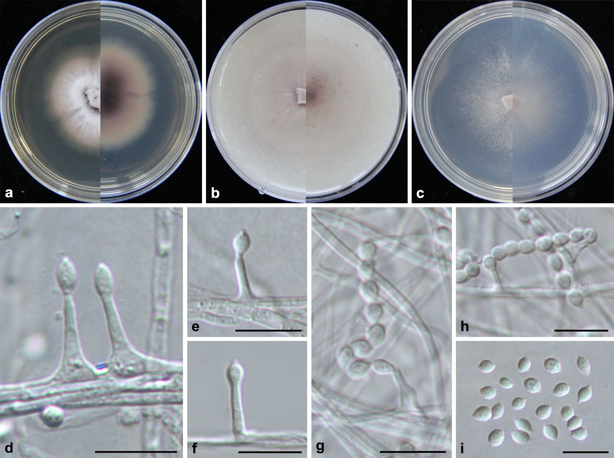
Aspergillus limoniformis (from ex-holotype CGMCC3.19323). a–c Upper and reverse views of cultures on PDA, OA and SNA 4 weeks after inoculation; d–h phialides and conidia; i conidia. Scale bars: d–i 10 µm