Aspergillus phialiformis Z.F. Zhang & L. Cai, sp. nov. 2020
Index Fungorum number: 556395; Facesoffungi number: FoF 08428
Holotype: CHINA, Yunnan, Yiliang, Sanjiao Cave, N 25.134°, E 103.383°, on rock, May 2016, Z.F. Zhang, HMAS 248017 (holotype designated here), ex-type living culture CGMCC3.19314 = LC12536; ibid., LC12537.
Morphological description
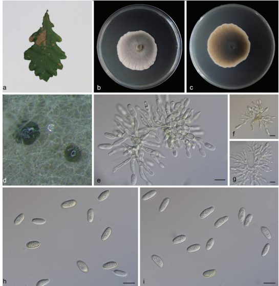
Hyphae hyaline, septate, smooth, branched, 1.0–2.5 μm wide. Asexual morph Conidiogenous cells simple phialides arising laterally on vegetative hyphae. Phialides cylindrical or tapering with enlarged base, occasionally branched, smooth, hyaline, variable in length, 4.0–12.0 µm long, 1.0–4.0 µm diam at base, tapering to 1.0–2.0 µm diam. at apex. Conidia formed in long chains, limoniform, subglobose or globose, apiculate, thick-walled, rough initially, then becoming smooth with age, hyaline, 2.5–4.0 µm ( x̄ ± SD = 3.3 ± 0.28, n = 60). Sexual morph not observed.Culture characteristics—Colonies on PDA attaining 36–41 mm diam. after 4 weeks, flat, margin fimbriate, cream yellow (4A2) at fruiting region, white to pale brown (5A2) from middle to aging region, with brown, radially striate and lobate ring, aerial mycelia sparse. Reverse cream-yellow (4A2) to brown (5C7). Colonies on OA attaining 31–36 mm diam. after 4 weeks, flat, margin undulate, aerial mycelia sparse, pulverulent in center, white. Reverse floralwhite (4A2). Colonies on SNA attaining 43–47 mm diam. after 4 weeks, flat, pulverulent, white. Reverse white. Sporulation within 3 weeks.
Habitat: on rock.
Distribution: Yunnan, Yiliang, Sanjiao Cave, China.
GenBank Accession:
Notes: Aspergillus phialiformis is phylogenetically closely related to A. phialosimplex (Fig. 9). While, phialides of A. phialiformis are cylindrical or basal enlarged, which are mostly cylindrical in A. phialosimplex. Meanwhile, limoniform conidia are not observed in A. phialosimplex and color of A. phialosimplex and A. phialiformis on PDA and OA are different.
Reference: Zhi‑Feng Zhang ,Shi‑Yue Zhou ,Lily Eurwilaichitret al.
Aspergillus phialiformis (from ex-holotype CGMCC3.19314). a–c Upper and reverse views of cultures on PDA, OA and SNA 4 weeks after inoculation; d–i phialides and conidia; j conidia. Scale bars: d–j 10 µm