Phanerochaete subrosea Y.L. Xu & S.H. He, sp. nov.2020
MycoBank: MB 835452; Facesoffungi number: FoF 08029
Holotype China, Ningxia Autonomous Region, Jingyuan County, Liupanshan Forest Park, on fallen angiosperm branch, 4 August 2015, He 2421 (BJFC 020874, holotype).
Morphological description
Fruiting body – Basidiomata annual, resupinate, effused, loosely adnate, easily detached from substrate, membranaceous, fragile after dried, up to 20 cm long, 3.5 cm wide. Hymenophore smooth, pale red (8A3), pastel red [8A(4–5)] to greyish red [8B(4–5)], turning purple in KOH, uncracked; margin thinning out, fimbriate or indistinct, lighter or concolorous with hymenphore surface; hyphal cords concolorous with hymenophore, turning purple in KOH.
Microscopic structures – Hyphal system monomitic; generative hyphae mostly simple-septate, occasionally with single or double clamp connections. Subicular hyphae colorless, slightly thickwalled, moderately branched and septate, tightly interwoven, more or less parallel to substrate, 3–5 µm in diam. Cystidia (leptocystidia) subcylindrical, colorless, thin-walled, smooth, with a basal simple septum, projecting above the hymenium, 33–55 × 3–5 µm. Basidia clavate, colorless, thinwalled, with a basal simple septum and four sterigmata, 24–36 × 4.5–5.5 µm; basidioles numerous, similar to basidia but smaller. Basidiospores ellipsoid, colorless, thin-walled, smooth, IKI–, CB–, 5–6 (–6.5) × 2.5–3 µm, L = 5.3 µm, W = 2.8 µm, Q = 1.9 (n = 30/1)
Habitat: on fallen angiosperm branch
Distribution: Ningxia Autonomous Region, northwest China.
GenBank Accession:ITS MT235687 nLSU MT248174 Literature This study
Notes: Phanerochaete subrosea is characterized by the light pink to red basidiomata turning purple in KOH and subcylindrical leptocystidia. Morphologically, the P. sanguinea group resembles P. subrosea by sharing the more or less red basidiomata with hyphal cords and presence of leptocystidia, but differs in turning black in KOH and staining the substrate with reddish orange color (Burdsall 1985, Bernicchia & Gorjón 2010, Floudas & Hibbett 2015). Phanerochaete carnosa is also similar to P. subrosea, but differs in having basidiomata turning green in KOH, subulate cystidia and slightly smaller basidiospores (Burdsall 1985). In the phylogenetic tree, P. subrosea is not closely related to the P. sanguinea group and P. carnosa, but forms a lineage with a sequence of P. krikophora (HHB-5796). However, this lineage received low support values from all analyses, and the latter name was not officially published and represented a species with clamp connections (Fig. 1, Floudas & Hibbett 2015). Phanerochaete subrosea resembles species of Rhizochaete Gresl., Nakasone & Rajchenb. by sharing the membranaceous basidiomata and hyphal cords turning purple in KOH, but they are not phylogenetically closely related (Greslebin et al. 2004).
Reference: Xu YL1 , Cao YF1 , Nakasone KK2 et al.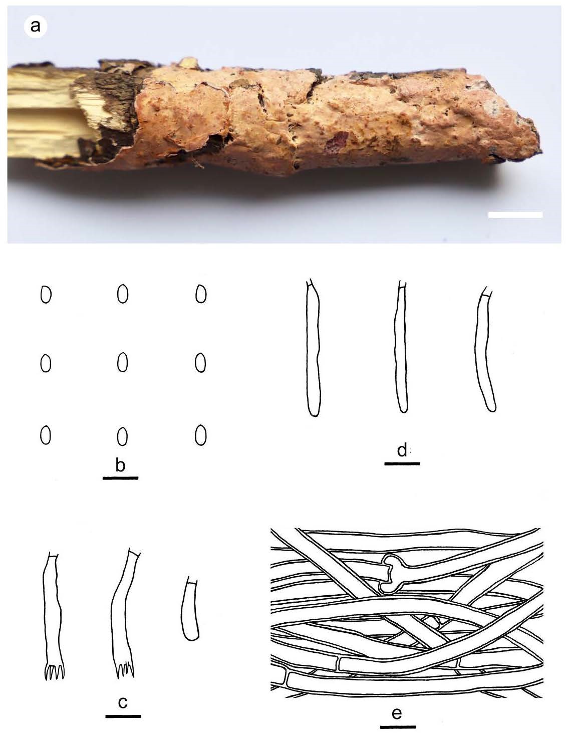
Figure 9 – Phanerochaete subrosea (from the holotype He 2421). a basidiomata. b basidiospores. c basidia and a basidiole. d cystidia. e. hyphae from subiculum. Scale bars: a = 1 cm, b–e = 10 µm