Phanerochaete sinensis Y.L. Xu, C.C. Chen & S.H. He, sp. nov. 2020
MycoBank: MB 835451; Facesoffungi number: FoF 08028
Holotype China, Liaoning Province, Zhuanghe County, Xianrendong Forest Park, on fallen angiosperm branch, 5 August 2017, He 4660 (BJFC 024179, holotype).
Morphological description
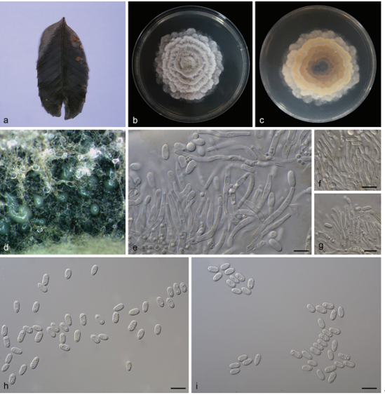
Fruiting body – Basidiomata annual, resupinate, effused, loosely adnate, easily detached from substrate, pellicular to membranaceous, up to 10 cm long, 4 cm wide. Hymenophore smooth, white (6A1), light orange (6A4) to greyish orange [6B(3–4)], slightly darkening in KOH, uncracked or sparsely cracked when dried; margin thinning out, fibrillose or indeterminate, concolorous with hymenophore surface; hyphal cords white (6A1) to orange [6B(7–8)], turning reddish brown in KOH.
Microscopic structures – Hyphal system monomitic; generative hyphae mostly simple-septate, occasionally with single or double clamp connections. Subicular hyphae colorless, slightly thickwalled, frequently branched and septate, loosely interwoven, more or less parallel to substrate, 3–7 µm in diam. Cystidia (leptocystidia) subcylindrical, slightly tapering toward apex, colorless, thinwalled, smooth, with a basal simple septum, projecting above the hymenium, 35–50 × 4–6 µm. Basidia clavate, colorless, thin-walled, with a basal simple septum and four sterigmata, 17–22 × 4– 5 µm; basidioles numerous, similar to basidia but smaller. Basidiospores ellipsoid to subcylindrical, colorless, thin-walled, smooth, IKI–, CB–, 4–5 (–5.5) × 2–2.5 µm, L = 4.9 µm, W = 2.2 µm, Q = 2.2 (n = 30/1).
Habitat: on fallen angiosperm branch
Distribution: Liaoning, Yunnan and Taiwan Provinces of China.
GenBank Accession:ITSMT235688\MT235689 nLSU MT248175\MT248176Literature This study\This study
Notes: Phanerochaete sinensis is characterized by the pellicular basidiomata with hyphal cords, presence of leptocystidia and subcylindrical basidiospores. It belongs to the P. burtii group, and morphologically almost indistinguishable from P. burtii, which, however, has longer basidia (25–35 µm) and a distribution outside China. For other comparisons with similar species, see the discussions under P. leptocystidiata above.
.
Reference: Xu YL1 , Cao YF1 , Nakasone KK2 et al.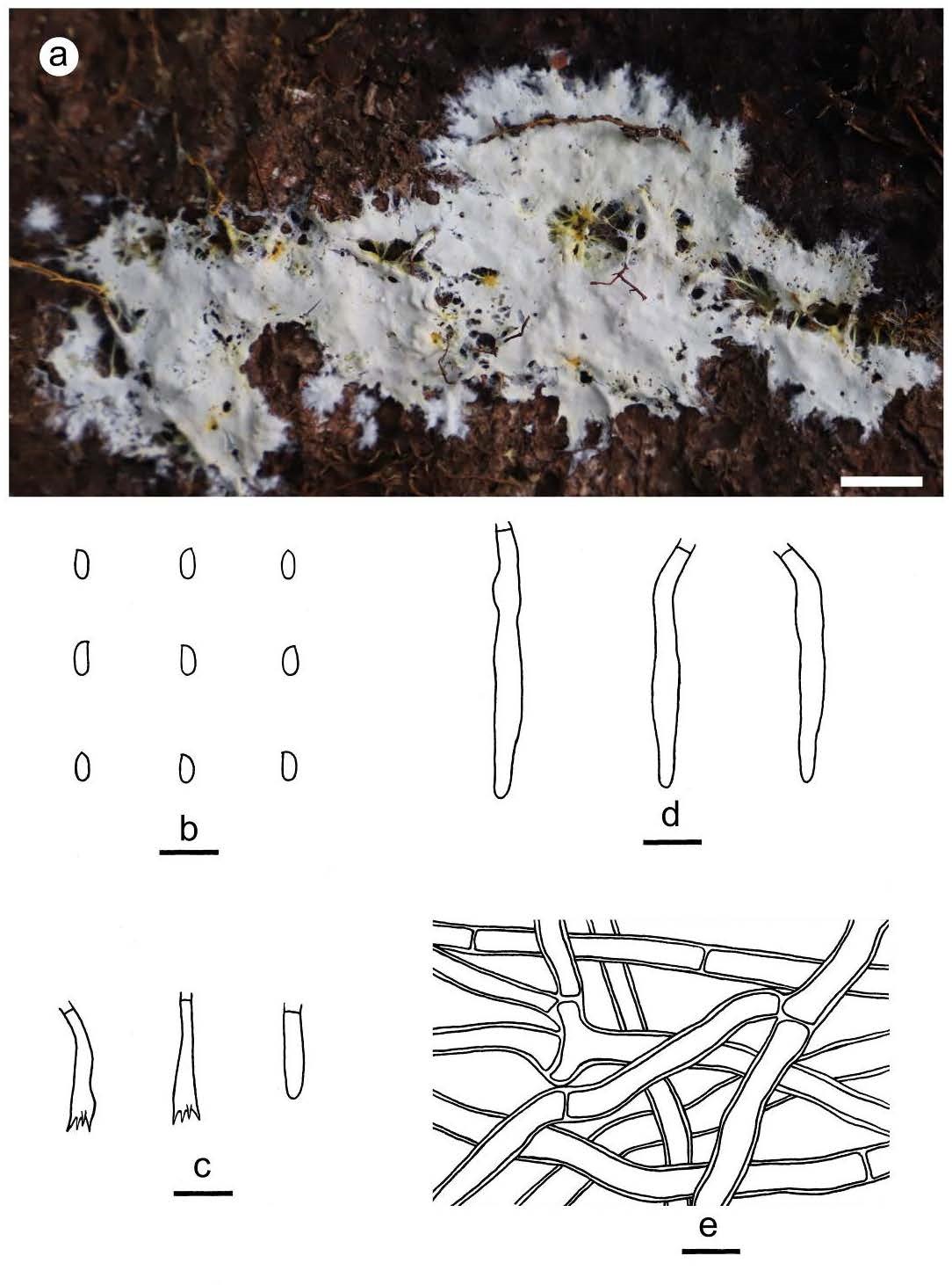
Figure 8 – Phanerochaete sinensis (a from He 6189, b–e from the holotype He 4660). a basidiomata; b. basidiospores; c. basidia and a basidiole; d. cystidia; e. hyphae from subiculum. Scale bars: a = 1 cm, b–e = 10 µm