Phanerochaete yunnanensis Y.L. Xu & S.H. He, sp. nov. 2020
MycoBank: MB 835448; Facesoffungi number: FoF 08030
Holotype China, Yunnan Province, Yongde County, Daxueshan Nature Reserve, on dead liana, 27 August 2015, He 2719 (BJFC 021157, holotype).
Morphological description
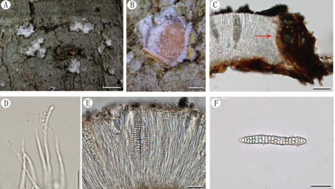
Fruiting body – Basidiomata annual, resupinate, effused, closely adnate, membranaceous to coriaceous, up to 20 cm long, 2.5 cm wide. Hymenophore grandinioid with small dense granules, pale orange (6A3), light orange [6A(4–5)] to greyish orange [6B(3–6)], unchanged in KOH, uncracked at first then densely cracked with age; margin thinning out, distinct, white, byssoid, becoming thick and indistinct with age.
Microscopic structures – Hyphal system monomitic; generative hyphae mostly simple-septate, occasionally with single or double clamp connections. Subicular hyphae colorless, thick-walled, moderately branched and septate, interwoven, more or less parallel to substrate, 3–5 µm in diam. Cystidia absent. Encrusted hyphae present, cylindrical, projecting above the hymenium, more or less grouped together. Basidia clavate to subcylindrical, colorless, thin-walled, with a basal simple septum and four sterigmata, 15–35 × 3.5–5 µm; basidioles numerous, similar to basidia but smaller. Basidiospores ellipsoid to subcylindrical, colorless, thin-walled, smooth, IKI–, CB–, 4.5–6 × 2–3 µm, L = 5.6 µm, W = 2.5 µm, Q = 2.2 (n = 30/1).
Habitat: on dead liana
Distribution: Yunnan Province, southwest China.
GenBank Accession: ITSMT235683 nLSU MT248166\MT248165 Literature This study\ This study
Notes: Phanerochaete yunnanensis is characterized by the light yellow grandinioid basidiomata, encrusted hyphae in hymenium, and absence of cystidia. Phanerochaete granulata Sheng H. Wu from Taiwan is similar to P. yunnanensis by sharing grandinioid basidiomata and absence of cystidia, but differs in lacking encrusted hyphae in hymenium and slightly shorter basidiospores (3.7–4.5 μm, Wu 2007). In addition, the ITS DNA sequences of type specimens of the two species are largely different (data not shown). Phanerochaete reflexa Sheng H. Wu is similar to P. yunnanensis by having uneven hymenophore and lacking cystidia, but differs in having tuberculate hymenophore with a reflexed margin (Wu 1998). Phanerochaete subquercina (Henn.) Hjortstam is similar to P. yunnanensis by sharing the odontioid hymenophore and absence of cystidia, but differs in having longer aculei and thin-walled hyphae, and lacks the encrusted hyphae in hymenium (Bernicchia & Gorjón 2010). Phanerochaete aculeata Hallenb. also has odontioid hymenophore, but differs from P. yunnanensis in having encrusted cystidia (Bernicchia & Gorjón 2010). In the phylogenetic tree, P. yunnanensis formed a sister lineage to P. robusta, but the latter can be easily distinguished by having smooth hymenophore and two kinds of cystidia (Wu et al. 2018b). Morphologically, P. yunnanensis resembles Hyphodermella corrugata (Fr.) J. Erikss. & Ryvarden, which also has grandinioid hymenophore and encrusted hyphae in hymenium, and lacks cystidia. However, they are not phylogenetically closely related.
.
Reference: Xu YL1 , Cao YF1 , Nakasone KK2 et al.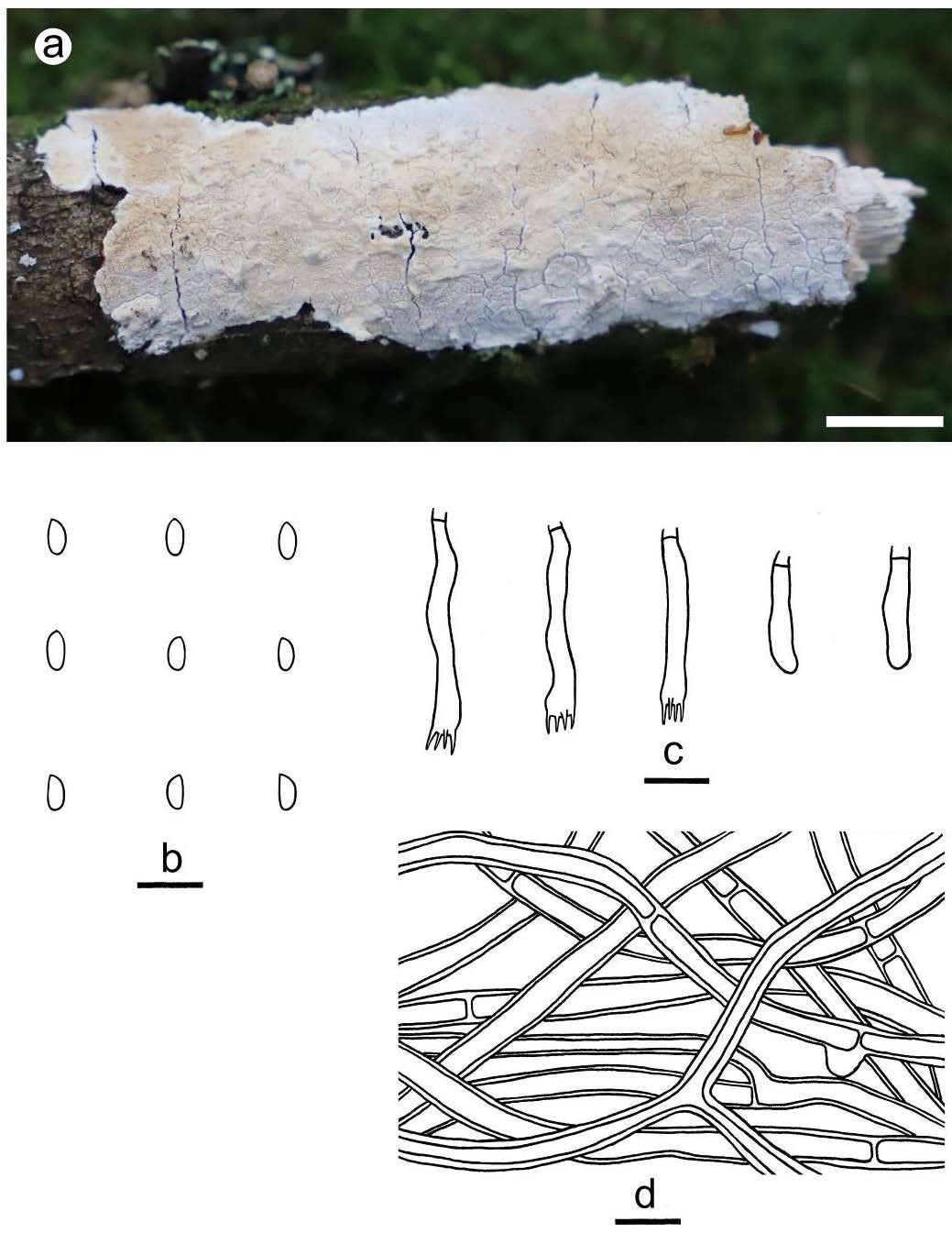
Figure 10 – Phanerochaete yunnanensis (a from He 6233; b–d from the holotype He 2719). a basidiomata. b basidiospores. c basidia and basidioles. d hyphae from subiculum. Scale bars: a = 1 cm, b–d = 10 µm.