Amphisphaeria yunnanensis L.S. Dissan., J.C. Kang & K.D. Hyde, sp. nov. 2020
Index Fungorum number: IF556876, Facesoffungi number: FoF 06505
Holotype: CHINA, Yunnan province, Qujing (24.668703oN, 104.24653oE), on a dead branch of an unknown host, 06 May 2019, L.S. Dissanayake, DW1137−048 (HMAS 290476, holotype; HKAS 107066, isotype), ex-type living culture KUMCC 19−0188, CGMCC, additional materials DW1137−049 (HAMS 290477, HKAS 107067), living culture KUMCC 19−0189.
Morphological description
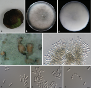
Saprobic on a dead branch. Sexual morph Ascomata 320–385 μm high × 380–450 μm diam. (x = 352.5 × 415 μm, n = 8), immersed, visible as black spots, solitary, scattered, globose to sub-globose, dark reddish brown, papillate. Ostiole 130−135 μm high × 28–32 μm diam. (x = 132.5 × 30 μm, n = 6). Peridium 8–18 μm (x = 13 μm, n = 10), comprising an inner layer of hyaline cells of textura angularis, and an outer layer of brown cells of textura angularis. Paraphyses 2–4.5 μm wide (x = 3.2 μm, n = 10), hyaline, few, longer than asci, cellular, constricted septate, guttulate, embedded in a gelatinous matrix, un-branched. Asci 78–93 × 6–9 μm (x = 85.5 × 7.5μm,n=20), 8-spored, unitunicate, cylindrical, with short pedicel, apically rounded, with a J- apical ring. Ascospores 12–15 × 4–6 μm (x = 13.5 × 5 μm, n = 30), overlapping uniseriate, fusiform, guttulate, hyaline when young, brown at maturity, uniseptate, constricted at the septum, smooth-walled. Asexual morph Undetermined. Culture characteristics:—Colonies on PDA, reaching 21.5 mm diam., after 2 weeks at 20–25 oC, medium dense, circular to slightly irregular with uneven margin, slightly raised and cottony surface, colony from above: white to pale grey at the margin, greenish-grey at the center; from below: yellowish white at the margin, yellow to brown at the center; mycelium greenish-grey.
Habitat: on a dead branch of an unknown host
Distribution: Yunnan Province, China
GenBank Accession: LSU MN556306,MN550992; ITS MN477177,MN550997
Notes: Both of our specimens (HMAS 290476 and HAMS 290477) share similar characters typical of Amphisphaeria species in having immersed ascomata, 8-spored, unitunicate asci and overlapping uniseriate, brown, uniseptate ascospores. Amphisphaeria yunnanensis is morphologically similar to A. bertiana Fairm., A. paedida (Berk. & Broome), Sacc. and A. vibratilis (Fuckel) E. Mull in having J- apical rings. However, Amphisphaeria bertiana has erumpent or superficial ascomata on a subiculum and A. paedida has superficial, coriaceous ascomata, while A. yunnanensis has immersed ascomata without a subiculum. Amphisphaeria vibratilis comprises with textura intricata cells in the peridium and larger ascospores (22.5–27.5 × 6–8.5 μm) with a mucilaginous sheath, while A. yunnanensis has compressed cells of textura angularis in the peridium and smaller ascospores (12–15 × 4–6 μm) without a sheath. Amphisphaeria yunnanensis has a close phylogenetic relationship with A. thailandica, but this is not supported in all formats of the analyses. However, A. yunnanensis differs from A. thailandica in having globose to sub-globose, dark reddish brown ascomata, long and narrow ostiole, fusiform, multi-guttulate ascospores. Moreover, A. yunnanensis differs from other remaining Amphisphaeria species in having narrow and long ostioles and small, fusiform, multiguttulate ascospores. Significant characteristics among similar taxa are given TABLE 2. Based on phylogenetic evidences and morphological differences, we introduce our new collection as a new species as A. yunnanensis.
Reference: LAKMALI S. DISSANAYAKE1,7, MILAN C. SAMARAKOON2,3,8, PETER E. MORTIMER4,9 et al.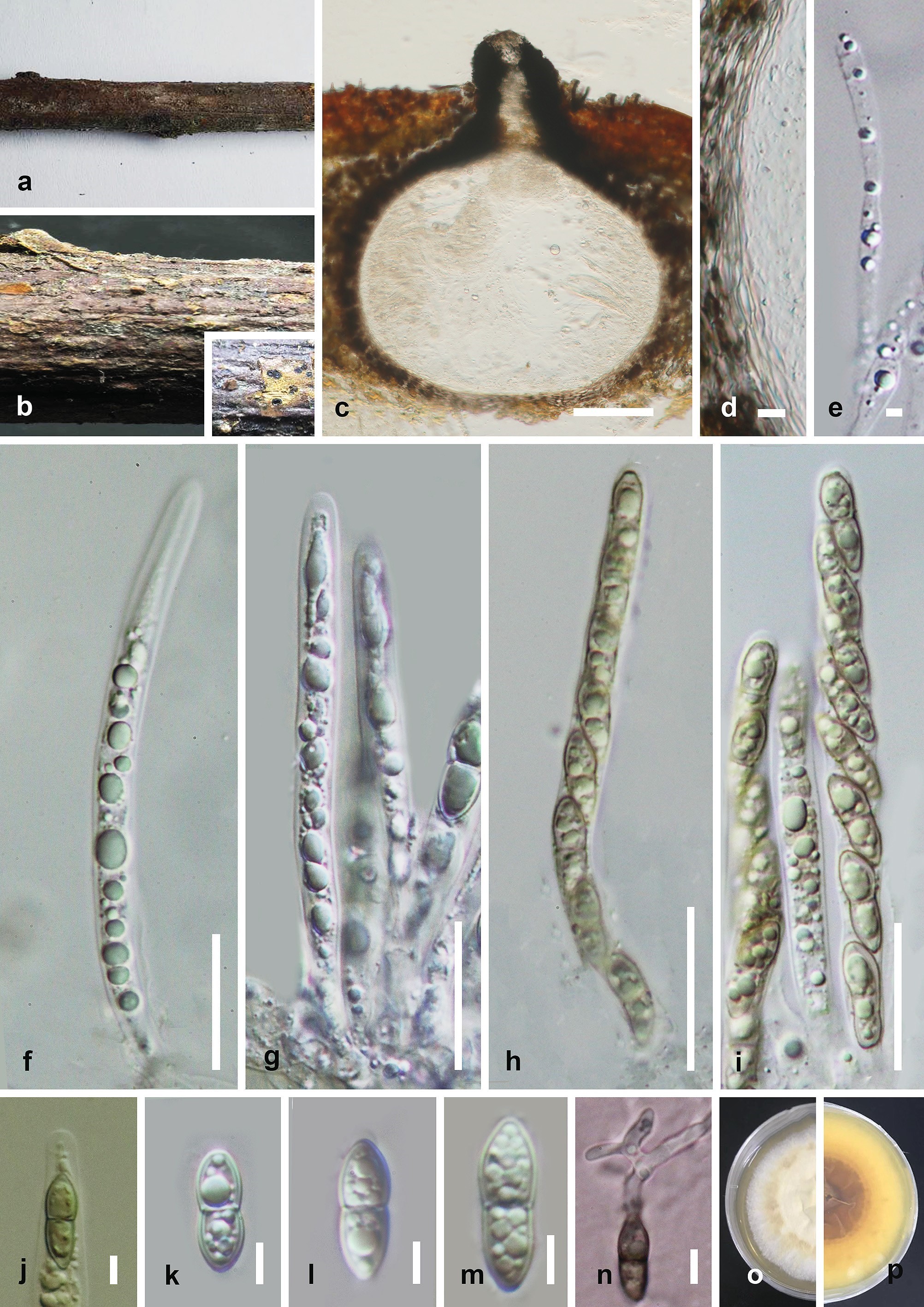
Amphisphaeria yunnanensis (HMAS 290476). a, b. Ascomata on the substrate. c. Vertical section of ascoma. d. Peridium. e. Paraphyses. f−i. Asci. j. J- Apical apparatus. k−m. Ascospores. n. Germinating ascospore. Culture on PDA from, o. above, p. below after 6 weeks. Scale bars: c = 100 μm, f−i = 20 μm, d,e, j−n = 5 μm.