Diaporthe minima Y.Y. Chen, A.J. Dissanayake and Jian K. Liu sp. nov. 2020
Index Fungorum number: IF557393; Facesoffungi Number: FoF07858; MycoBank Number: MB836215
Holotype: China, Guizhou Province, Guiyang District, Huaxi Wetland Park, Saprobic on decaying branch, April 2017, Y.Y. Chen (HKAS 107539, holotype), ex-type living culture CGMCC 3.20097 = GZCC 19-0066; ibid., (GZAAS 19-1786, paratype), living culture GZCC19-0070; ibid., GZAAS 19-1787, living culture GZCC19-0061; ibid., GZAAS 19-1788, living culture GZCC19-0207.
Morphological description
Saprobic on decaying woody branch. Sexual morph: Not observed. Asexual morph: Conidiomata up to 230 µm in diam., immersed, scattered on PDA, dark brown to black, globose, solitary or clustered in groups of 3–5 conidiomata. Conidiophores 9–13 × 1–2 µm (x = 11 × 1.5 µm) aseptate, cylindrical, straight or sinuous, densely aggregated, terminal, slightly tapered towards the apex. Alpha conidia 6.5–8.5 × 2–3 µm (x = 7 × 2 µm), biguttulate, hyaline, fusiform or oval, both ends obtuse. Beta conidia not observed.Culture characteristics: Cultures incubated on PDA at 25 ◦C in darkness. Colony at first flat with white felty mycelium, becoming black in the center and black at the marginal area with 8 d, pycnidia not observed.
Habitat: on decaying branch
Distribution: China
GenBank Accession:
Notes: The phylogenetic result showed that isolates of Diaporthe minima clustered closer toD. bohemiae, D. juglandicola and D. rostrata, and formed a distinct lineage (Figure 1) with maximum support (ML/MP/BI = 100/100/1.0). Diaporthe minima can be distinguished from the above closely related species based on ITS, tef, tub, cal and his loci for D. bohemiae (11/539 in ITS, 45/467 in tef, 14/453 in tub, 37/606 in cal, 34/513 in his), D. juglandicola (24/539 in ITS, 19/467 in tef, 12/453 in tub, 27/606 in cal and 47/513 in his) and D. rostrata (19/539 in ITS, 48/467 in tef, 13/453 in tub 17/606 in cal and 49/513 in his). Morphologically, Diaporthe minima differs from D. bohemiae, D. juglandicola and D. rostrata in having smaller alpha conidia (7 × 2 vs. 9 × 3 µm) (7 × 2 vs. 11 × 13 µm) [17,18].
Reference: Asha J. Dissanayake, Ya-Ya Chen and Jian-Kui (Jack) Liu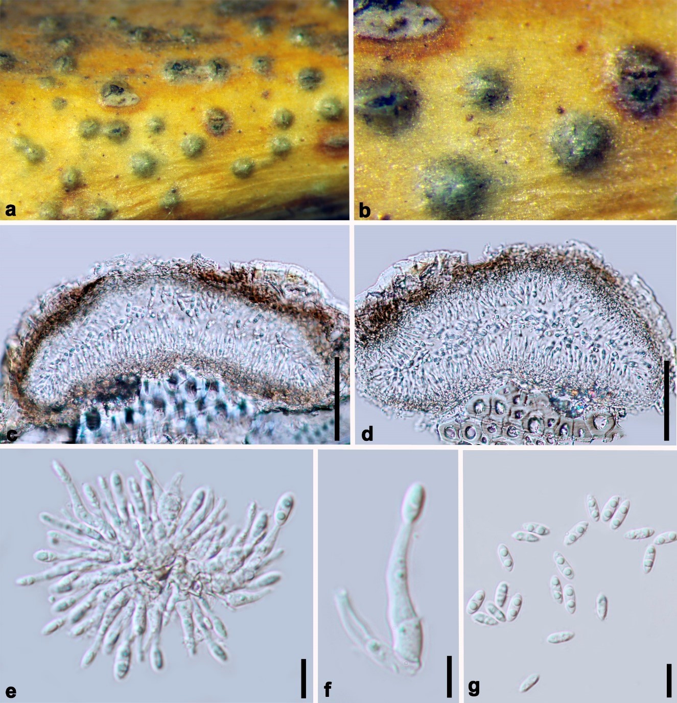
Diaporthe minima (HKAS 107539, holotype). (a,b) Conidiomata on host surface. (c,d) Section of conidiomata. (e,f) Alpha conidia attached to conidiogenous cells. (g) Alpha conidia. Scale bars: (c,d) = 50 µm, (e–g) = 10 µm.