Diaporthe minusculata Y.Y. Chen, A.J. Dissanayake and Jian K. Liu sp. nov. 2020
Index Fungorum number: IF557394; Facesoffungi Number: FoF07859; MycoBank Number: MB836216
Holotype: China, Guizhou Province, Xingyi Wanfenglin, Saprobic on decaying branch,
June 2019, Y.Y. Chen (HKAS 107540, holotype), ex-type living culture CGMCC 3.20098 = GZCC 19-0215; ibid., GZAAS 19-2072, living culture GZCC 19-0372; ibid., Suiyang broad water nature reserve, saprobic on decaying woody host, April 2018, Y. Y. Chen (GZAAS19-2062, paratype), living culture GZAAS19-2062; ibid., GZAAS19-2070, living culture GZCC 19-0366.
Morphological description
Saprobic on decaying branch. Sexual morph: Not observed. Asexual morph: Conidiomata up to 430 µm in diam., superficial, erumpent, scattered on PDA, dark brown to black, globose, solitary or clustered in groups of 3–5 pycnidia, with prominent necks 130–320 µm long. Conidiophores 11–18 × 1.5–2.5 µm (x = 14 × 2 µm), aseptate, cylindrical, straight or sinuous, densely aggregated, terminal, slightly tapered towards the apex. Alpha conidia 7–10 × 2–3 µm (x = 9 × 2 µm), biguttulate, hyaline, fusiform or oval, both ends obtuse. Beta conidia not observed.Culture characteristics: Cultures incubated on PDA at 25 ◦C in darkness showed colony at first white, becoming pale brown with yellowish dots within 10 d, with dense and felted mycelium, visible solitary or aggregated pycnidia at maturity.
Habitat: on decaying branch
Distribution: China
GenBank Accession:
Notes: The phylogenetic results showe that Diaporthe minusculata clustered close to D. malorum and D. passiflorae, and formed a distinct lineage (Figure 1) with maximum support (ML/MP/BI =100/100/1.0). Diaporthe minusculata can be distinguished from D. malorum (23/539 in ITS, 41/467 in tef, 29/453 in tub, 52/606 in cal and 35/513 in his) and from D. passiflorae (25/539 in ITS, 13/467 in tef, 11/453 in tub, 22/606 in cal and 17/513 in his). Morphologically, Diaporthe minusculata differs from D. malorum in having conidiomata with a long neck and differs from D. passiflorae in shorter conidiophores (14 × 2 vs.26 × 4 µm) [16–18,66,74].
Reference: Asha J. Dissanayake, Ya-Ya Chen and Jian-Kui (Jack) Liu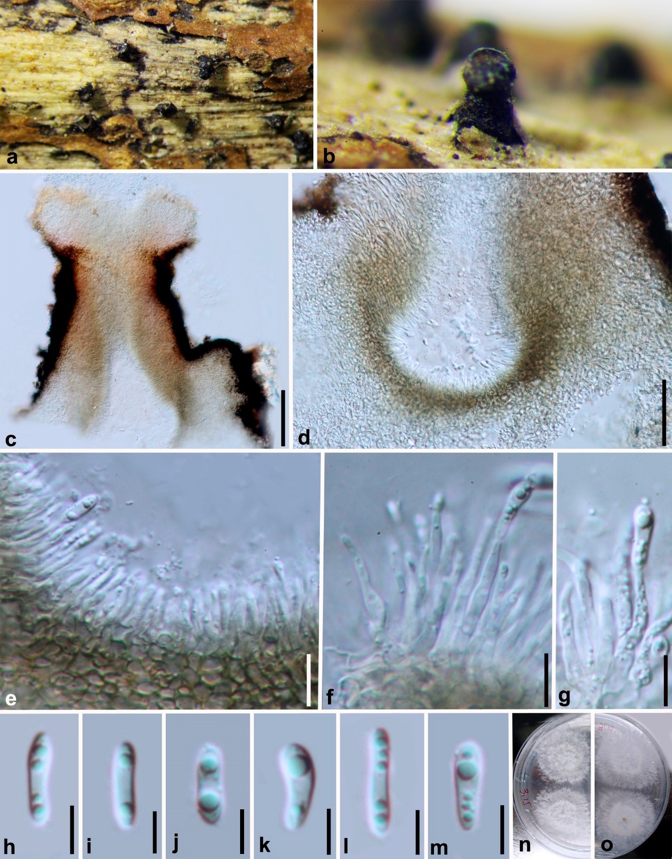
Diaporthe minusculata (HKAS 107540, holotype). (a,b) Conidiomata on host surface. (c–e) Section of conidiomata. (f,g) Alpha conidia attached to conidiogenous cells. (h–m) Alpha conidia.
(n) 5 days old culture on PDA from above. (o) 5 days old culture on PDA from reverse. Scale bars: (c) = 100 µm, (d) = 50 µm, (e–g) = 10 µm, (h–m) = 5 µm.