 83
83
Plectosphaerella endophytica Z.F. Yu & X.Q. Yang, sp. nov.2021
MycoBank No: 838656
Holotype: China, Yunnan Province, Kunming, The Dian Lake, 24°96'N, 102°66'E, 1886 m alt., isolated from Hydrilla verticillata (L.f.) Royle as an endophyte, 20 Jul. 2014, Z.F. Yu, YMF 1.04701 (Holotype), ex-type CCTCC AF 2021053.
Morphological description
Sexual morph: Undetermined
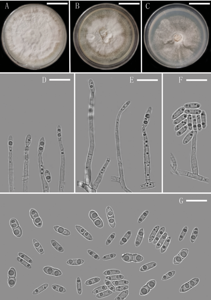
Asexual morph: Colony on CMA after 3 d, hyphae hyaline, smooth, septate, thin walled, branched, 1.9–3.3 µm ( = 2.6 µm, n = 10) wide. Conidiophores macronema tous, mononematous, erect, straight or flexuous, smooth-walled, hyaline, unbranched or occasionally irregular branched, sometimes 1–2-septate. Conidiogenous cells phi alides, subulate, integrated, terminal, determinate, hyaline, smooth-walled. Conidia solitary, acrogenous, broadly navicular to broadly fusiform, suboblong or ellipsoidal, 0–1-septate, usually constricted at septum, bi-guttulate, hyaline, smooth-walled, asep tate conidia abundant, 5–9.1 × 2.5–3.5 µm (= 7.8 × 3.1 µm, n = 30) ;septate conidia scarce ,8.8–10.1 × 3.7–4.6 µm ( = 9.4 × 4.1 µm, n = 30), forming hyaline to white mucilaginous masses. Sexual morph and chlamydospores absent.
Culture characteristics: Colonies on OA reaching 52 mm diameter, on PDA reaching 48 mm diameter and on CMA reaching 43 mm diameter in 14 d at 25 °C. On PDA, colonies white, dense, fluffy hyphae growth in the medium surface, outer most mycelia formed an annule, margin smooth and entire, sporulation abundant, reverse pale yellow to white.
Habitat:
Distribution: China
GenBank Accession: LSU MW024052; ITS MW024054; TEF-1α MW029607; TUB2 MW029608
Notes: Although the phylogenetic analyses showed that our isolate Plectosphaerella endophytica is close to P. oratosquillae, the conidia of P. oratosquillae are aseptate, multi guttulate (Duc et al. 2009). Furthermore, P. endophytica is most similar to P. verschoorii Hern.-Restr. & Giraldo López in the septa of conidia; both species produce 0–1-sep tate conidia, and septate conidia are larger than aseptate conidia (P. verschoorii: 1-sep tate conidia, 8–11.5 × 2–3 µm; aseptate conidia, 3–8.5 × 2–3 µm), but there are obvi ous difference in the shape of conidia, P. endophytica was deeply constricted at septa. Besides, the phialides of P. verschoorii are shorter (up to 14 µm) (Giraldo et al. 2019).
Reference: [1] Yang, X. , Ma, S. , Peng, Z. , Wang, Z. , Qiao, M. , & Yu, Z. . (2021). Diversity ofplectosphaerellawithin aquatic plants from southwest china, withp. endophyticaandp. sichuanensisspp. nov. MycoKeys, 80, 57-75.
Plectosphaerella endophytica (YMF 1.04701, holotype) A–C colony on OA, PDA and CMA after 14 d D–F conidiophores and Phialides G conidia. Scale bars: 1.35 cm (A–C), 10 µm (D–G).

