Trichoderma camelliae Jayaward & Manawas, sp. nov. 2021
MycoBank Index Fungorum: IF558000, Facesoffungi Number: FoF09390
Holotype: JZBH3360002.
Morphological description
Sexual morph:
Asexual morphs: Mycelia aseptate, branched, effused Conidiophores scattered, dark green to greyish–green, tree–like, comprising a main axis. Conidiogenous cells ampulliform, arising singly as clusters. Conidia developed at the hyphal end also observed. Conidia 1.5 –2× 1–2 μm (x̅= 2×2 μm, n = 40) ovoid to short ellipsoidal, verrucose.
Cultures: On PDA mycelium covers plate after three days at 25°C. Aerial hyphae, hairy dense internal zone, initially white mycelium with time become pale yellow. Develop abundant, and flat large green disc around the inoculum, turning green.
Habitat: on dead Camellia sinensis leaves and shoots.
Distribution: CHINA, Fujian Province, Zhangzhou County.
GenBank Accession: ITS: MW008451;Notes: The isolates obtained in the present study fit well morphologically within the Trichoderma. The present species, Trichoderma camelliae, developed a strongly supported monophyletic clade with 100% ML and 1.0 BYPP values. Morphologically this species differs from the type species of Trichoderma viride, by developing ellipsoidal and larger conidia, whereas conidia of the type species are mostly ovoid and smaller than the species identified in this study (0.7 µm long and 1 µm diam.) (Lieckfeldt et al. 1999).
Reference: Manawasinghe IS, Jayawardena RS, Li HL, Zhou YY, Zhang W, Phillips AJL, Wanasinghe DN, Dissanayake AJ, Li XH, Li YH, Hyde KD, Yan JY 2021 – Microfungi associated with Camellia sinensis: A case study of leaf and shoot necrosis on Tea in Fujian, China. Mycosphere 12(1), 430–518, Doi 10.5943/mycosphere/12/1/6
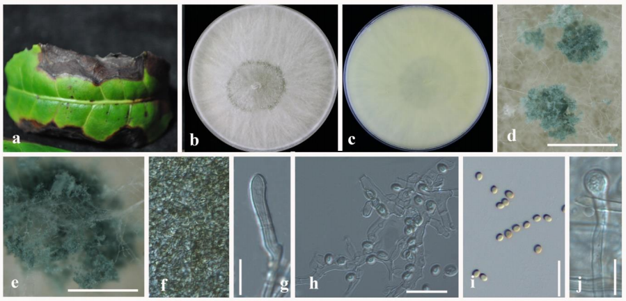
Trichoderma camelliae (JZB3360002 ex–holotype). a Diseased leaf. b Upper view of the colony on PDA after three days. c Reverse view of the colony on PDA after three days. d–e Conidiomata on PDA. f Pycnidal wall. g Conidiogenous cell. h–i Conidia. j Germinating conidium. Scale bars: d, e = 100 µm, f, g = 100 µm, h–j = 10 µm.