Botryosphaeria dothide Ces. & De Not. 2021
MycoBank Index Fungorum: IF 183247; Facesoffungi number: FoF03512
Holotype:
Morphological description
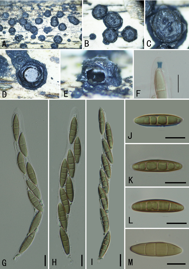
Sexual morph:
Asexual morphs: Conidiomata stromatic, Conidiophores hyaline, cylindrical, smooth. Conidiogenous cells 11.5–14 × 4–6.5 μm (x = 13 × 6 m, n = 20), hyaline, sub–cylindrical. Conidia 18–40 × 5–10 μm (x̅= 24 × 7 μm, n = 20), hyaline, unicellular, narrowly fusiform, with a sub–truncate to bluntly rounded base, forming a septum before germination, smooth–walled with granular contents.
Cultures: Colonies on PDA reaching 50 mm diam., after four days at 28°C. Initially, white becoming grey, moderately dense, margin smooth, olivaceous.
Habitat: on dead leaves and shoots of Camellia sinensis.
Distribution: CHINA, Fujian Province, Zhangzhou County.
GenBank Accession: ITS: MT497875;tub2: MT513138;tef1: MT513143.
Notes: The colony morphology of taxa isolated in this study are similar to typical strains of B. dothidea (Phillips et al. 2013). In the multilocus phylogenetic analysis, the five isolates from the present study clustered together with 56% ML bootstrap and less than 0.90 BYPP. Morphologically these taxa are similar to the type description of B. dothidea (Phillips et al. 2013). Botryosphaeria dothidea has a wide range of hosts and it is a well–known woody host–pathogen (Phillips et al. 2005, 2013, Dissanayake et al. 2016, Hyde et al. 2020a). Botryosphaeria dothidea had been reported to cause diseases on many different hosts in China (Manawasinghe et al. 2018). This species was first reported as shoot blight pathogen in Chinese tea plants in 2016 (Jayawardena et al. 2016b).
Reference: Manawasinghe IS, Jayawardena RS, Li HL, Zhou YY, Zhang W, Phillips AJL, Wanasinghe DN, Dissanayake AJ, Li XH, Li YH, Hyde KD, Yan JY 2021 – Microfungi associated with Camellia sinensis: A case study of leaf and shoot necrosis on Tea in Fujian, China. Mycosphere 12(1), 430–518, Doi 10.5943/mycosphere/12/1/6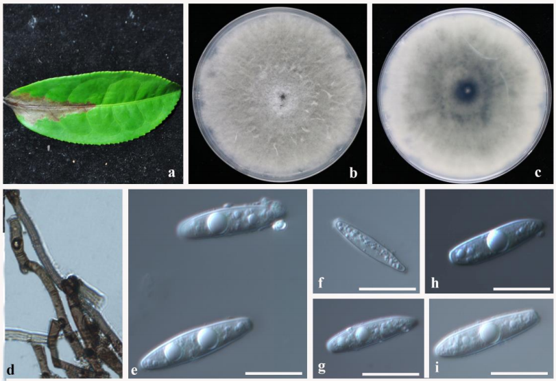
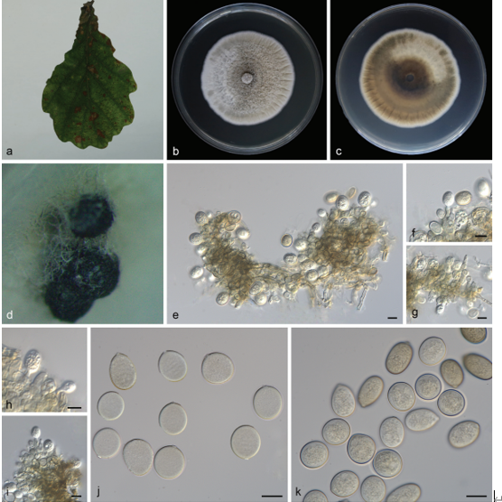
Botryosphaeria dothidea (JZB310193) a Material examined. b Upper view of the colony on PDA after four days. c Reverse view of the colony on PDA after four days. d Mycelia. e–i Conidia. Scale bars: e–i = 20 µm.