Phragmidium jiangxiense Y.M. Liang & Y. Liu 2020
MycoBank MB832198
Holotype: CHINA. JIANGXI: Qiyun Mountain, 114°1.8′E, 25°49.8′N, 681 m asl, on Rosa laevigata, 20 Jul 2018, Y.M. Liang (holotype BJFCR03453). GenBank: ITS = MN264715; 28S = MN264733.
Morphological description
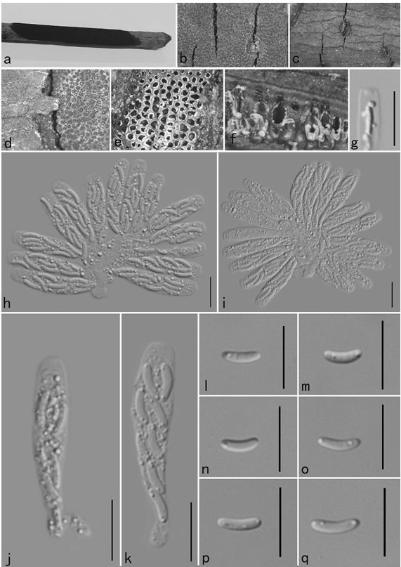
Spermogonia unknown. Aecia and uredinia produced together on the abaxial leaf surface, hypophyllous, scattered to gregarious, rounded, 0.2–0.5 mm diam, bright orange-colored, and powdery. Aeciospores produced in basipetal succession, subglobose, 13–22 × 12–17 μm, cytoplasm orange; wall 1–2 μm thick, colorless, echinulate on the smooth surface. Uredinia paraphysate; paraphyses clavate, thin-walled, 22–41 × 6–10 μm. Urediniospores produced singly on a pedicel, oval, 15–23 × 11–18 μm, cytoplasm orangecolored; wall 0.5–2 μm thick, colorless, echinulate with stout spines basally submersed in wrinkled spore wall and gradually disappearing toward the apical area. Teliospores not observed.
Habitat: On Rosa laevigata.
Distribution: In China.
GenBank Accession: ITS :MN264715; 28S :MN264733.
Notes: Phragmidium jiangxiense is sister to P. japonicum with MP/ML values of 69/52 (FIG. 1). However, the life cycle pattern distinguishes it from P. japonicum. Phragmidium jiangxiense is apparently macrocyclic, producing aecial and uredinial stages, whereas P. japonicum is microcyclic, as proven by experimental inoculations (Ono 2002). Rosa laevigata is also the host of P. rosae-multiflorae and P. tuberculatum (Tai 1979; Wei 1988; Hiratsuka and Chen 1991; Hiratsuka et al. 1992; Cho and Shin 2004; Zhuang et al. 2012). We observed many specimens of P. rosae-multiflorae and P. tuberculatum (SUPPLEMENTARY TABLE 1; Liu et al. 2018; Ono 2019). The three species differ among each other by the surface structure of their aeciospores. The aeciospore surface of P. rosae-multiflorae is echinulate above and becomes smooth toward the base, whereas that of P. tuberculatum is completely echinulate (Wahyuno et al. 2002; Liu et al. 2018; Ono 2019). The aeciospore surface of P. jiangxiense is echinulate with stout spines basally submersed in the wrinkled spore wall (FIG. 3). In addition, P. rosae-multiflorae and P. tuberculatum clustered apart distantly from P. jiangxiense (FIG. 1). The unique spore morphology and autonomous phylogenetic position support the conclusion that P. jiangxiense is a distinct species.
Reference: Yun Liua, Ying-Mei Liang b, and Yoshitaka Onoc

Phragmidium jiangxiense (BJFCR03452, holotype). A. Aecia and uredinia. B. Aeciospores, paraphyses, and urediniospores under light microscope. C. Aeciospores and urediniospores under light microscope. D. Aeciospores, paraphyses, and urediniospores under SEM. E–F. Urediniospores under SEM. G–J. Urediniospores under SEM. Bars: B–E, G–I = 5 µm; F, J = 1 µm.