Phragmidium leucoaecium Y.M. Liang & Y. Liu 2020
MycoBank MB832100
Holotype: CHINA. YUNNAN: Jizu Mountain, Binchuan, Dali, 100°23.6′E, 25°57.6′N, 2245 m asl, on Rosa sp., 22 Mar 2015, Y.M. Liang & B. Cao (holotype BJFCR02118). GenBank: ITS = MN264719; 28S = MN264737.
Morphological description
Spermogonia epiphyllous, subepidermal, of determinate growth, dome-shaped with flat hymenium (type 2), 103–220 μm wide and 55–70 μm high. Aecia hypophyllous, subepidermal, break through the epidermis at later period, spreading up to 12 mm long, often becoming round white spots, aparaphysate, white or infrequently dull yellowish, powdery. Aeciospores produced in basipetal succession, ellipsoid to broadly ellipsoid, generally angular, 24–40 × 12–22 μm, cytoplasm colorless; wall 1–2 μm thick, colorless, covered with irregularly elongated verrucae at the base and conical projections interconnected by narrow ridges toward the apex.
Habitat: On Rosa sp.
Distribution: In China.
GenBank Accession: ITS : MN264719; 28S :MN264737
Notes: The spermogonia of Phragmidium leucoaecium are type 2, which is common in the families Pucciniastraceae Gäum. & Leppik and Melampsoraceae Dietel (Cummins and Hiratsuka 2003). Type 2 spermogonia were also found in P. warburgianum (syn. Kuehneola warburgiana; Ono 2012). Phragmidium leucoaecium and P. warburgianum are distinct from each other in the surface structure of aeciospores. The surface of the aeciospores in P. leucoaecium is covered with irregularly elongated verrucae at the base and conical projections interconnected by narrow ridges toward the apex, whereas that of P. warburgianum is covered with regular circular verrucae. The wall surface structure of aeciospores of P. leucoaecium is different from other Phragmidium species that parasitizes Rosa plants. The unique aeciospore surface structure and an autonomous phylogenetic position support P. leucoaecium as a species distinct from all known Phragmidium species.
Reference: Yun Liua, Ying-Mei Liang b, and Yoshitaka Onoc
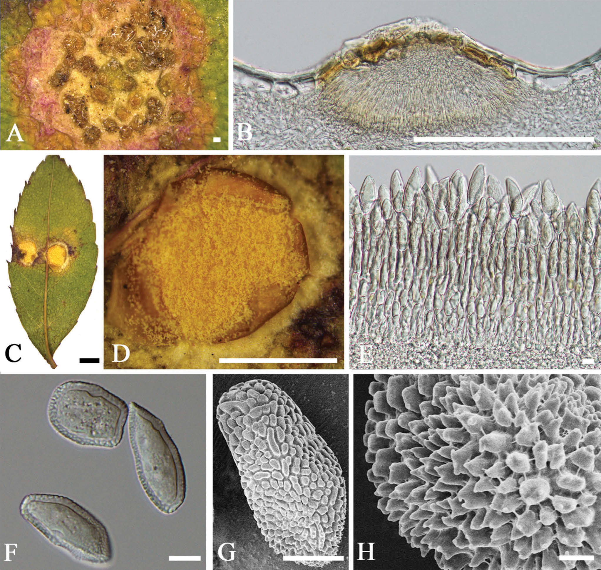
Phragmidium leucoaecium (BJFCR02118, holotype). A–B. Spermogonia. C–D. Aecia. E–F. Aeciospores under light microscope. G–H. Aeciospores under scanning electron microscope. Bars: A, B = 100 µm; C, D = 1 cm; E–G = 10 µm; H = 1 µm.