Gymnosporangium tiankengense P. Zhao & L. Cai 2020
MycoBank MB832746
Holotype: China, Guangxi, Bai Se, Leye County, Shenmu Tiankeng, 0, I on Malus sp., 18 June 2017, P. Zhao (holotype HMAS248124). SSU, ITS and LSU sequences GenBank MN605026, MN605725 and MN605803.
Morphological description
Spermogonia, uredinia and telia not found. Aecia foliicolous, hypophyllous, roestelioid; peridium tubular, spreading or erect, 3–8.5 mm high, peridial cells rhomboid-oblong, 34–77 × 17–32 mm, outer walls smooth, inner walls small papillae and side walls moderately rugose; aeciospores globoid, ovoid, large coronate, 16–24 × 14–20 mm, walls yellowish, 1.0–2.0 mm thick, germ pore 3–7, scattered.
Habitat: On Malus sp.
Distribution: In China.
GenBank Accession:
Notes: This species is characterised by its relatively smaller peridial cells and aeciospores. Aecial morphological differences of this rust were distinct from other Gymnosporangium species on Malus species, except G. libocedri (Kern 1973). Based on our morphological comparison, G. libocedri has relatively short peridia on aecia, and large peridial cells(77–148 × 15–29 μm), which clearly differs from our newly proposed species. The ornamentation of aeciospores in G. libocedri is verrucose with refractive granules and its peridial cells with verrucose outer walls and inner walls. In addition, this new species resembles to G. shennongjiaense but differs in length of peridia, dimension of peridial cells and aeciospores. These morphological characters can clearly differentiate the two species, and molecular data further supported the phylogenetic distinction of this species from other Gymnosporangium species. This species was discovered at the edges of Tiankeng in Guangxi province in southwest part of China.
Reference: P. Zhao1, X.H. Qi 2, P.W. Crous3,4 et al.
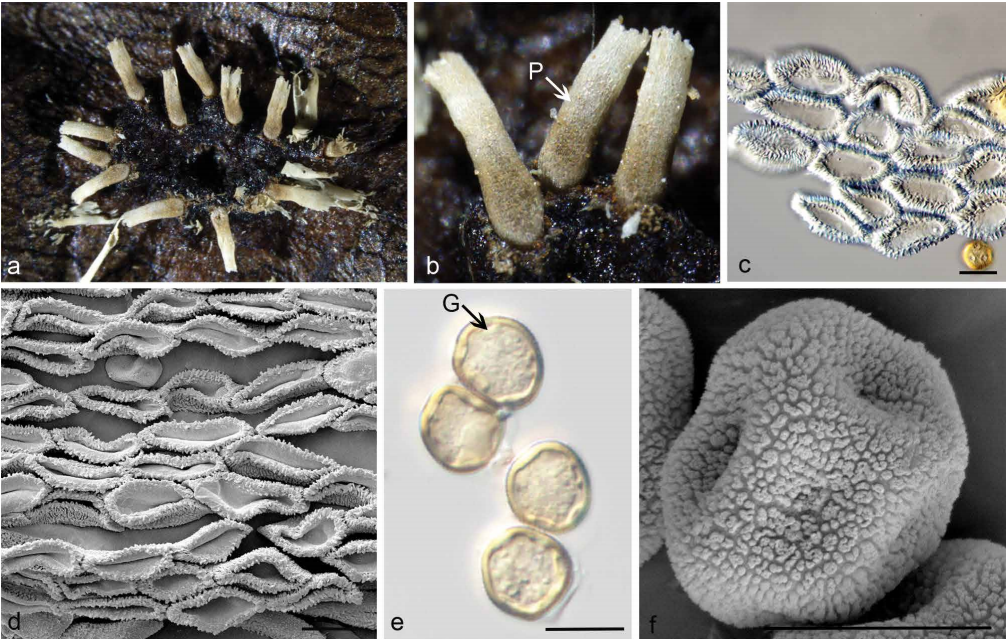
Morphology of G. tiankengense. a. Aecia on the hypophyllous leaf surface; b. erect peridia (P) on the hypophyllous leaf surface; c. oblong peridial cells observed by LM; d. ultrastructure of peridial cells observed by SEM; e. globoid or ellipsoid aeciospores with scattered germ pores (G) observed by LM; f. verrucose aeciospores observed by SEM. — Scale bars: c–f = 20 µm.