Gymnosporangium spinulosum P. Zhao & L. Cai 2020
MycoBank MB832745
Holotype: China, Sichuan, Chengdu, 0, I on M. spectabilis, 19 May 1955, Y.C. Wang (holotype HMAS26416). SSU, ITS and LSU sequences GenBank MN605030, MN605727 and MN605805.
Morphological description
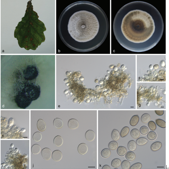
Spermogonia, uredinia and telia not found. Aecia foliicolous, hypo- phyllous, roestelioid, 4–9 mm high, tubular; peridium cornuted, rupturing at apex, peridial cells rhomboid or oblong, 55–86 × 14–23 μm, outer cells rugose, inner walls densely verrucose with long papillae up to 5 μm; aeciospores globoid, 14–25 × 13–22 μm, walls slightly brown, 2 μm thick, evenly thickened, walls large coronate, basal parts columnar and upper parts separated into several long protuberances.
Habitat: On M. spectabilis.
Distribution: In China.
GenBank Accession:
Notes: This species is characterised by its special ornamentation in peridial cells and aeciospores. In addition, it has relatively small aeciospores. Compared to other Gymnosporangium species, it resembles G. asiaticum in the dimension of its aeciospores, and the two species have commonly been confused in the past (Wang & Guo 1985, Zhuang 2012). How- ever, these two species clearly differ in ornamentation of peridial cells and aeciospores. This novel species has peridial cells with papillae up to 5 μm long, which clearly differs from those in G. asiaticum. Besides, it has aeciospores with basal parts columnar and upper parts separated into several long protuberances, and this character can clearly differentiate the two species. Phylogenetic results further supported this species distinct from G. asiaticum and other species.
Reference: P. Zhao1, X.H. Qi 2, P.W. Crous3,4 et al.
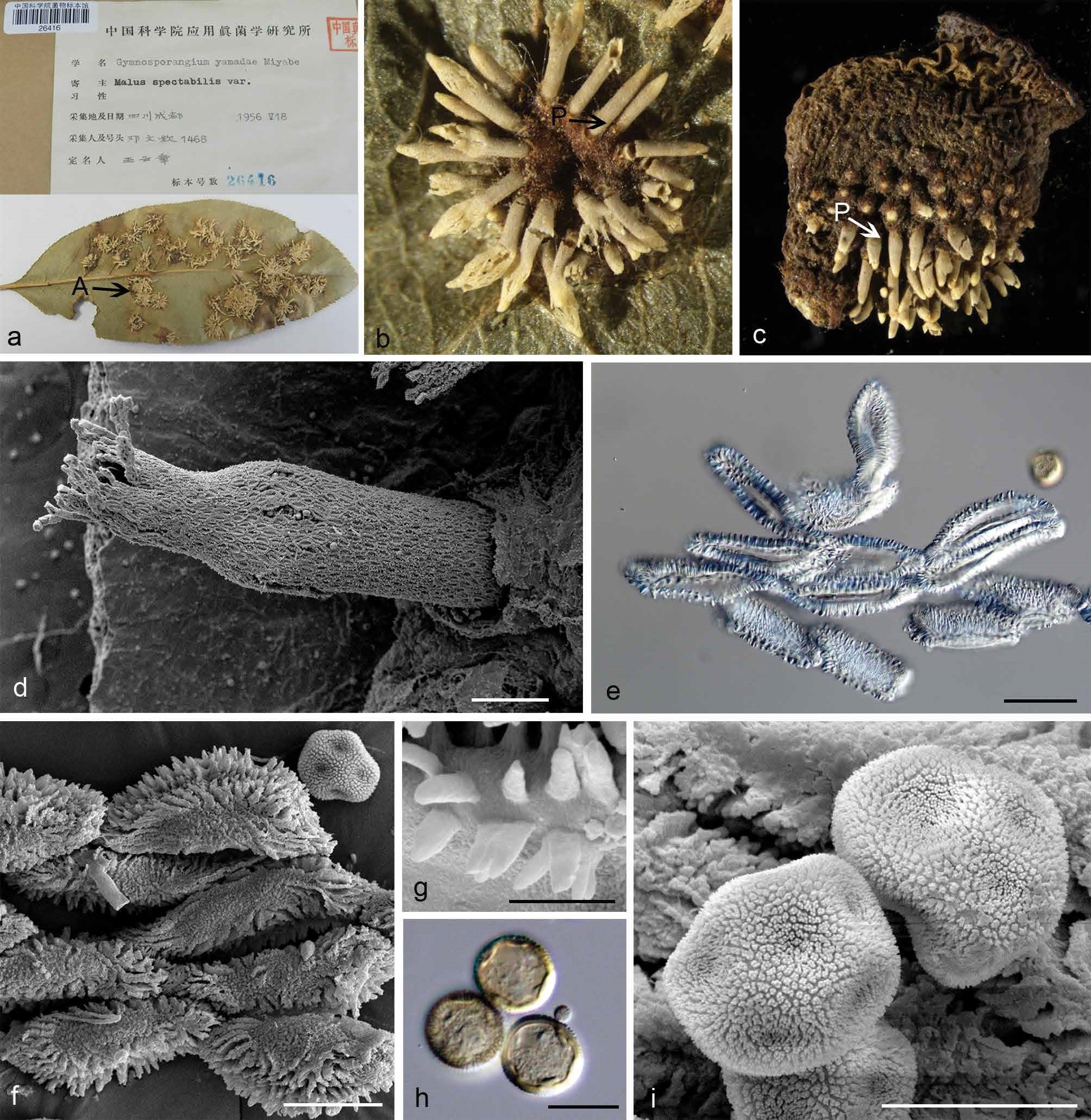
Morphology of G. spinulosum. a. Labels of the holotype specimen and aecia (A) on the hypophyllous leaf surface; b. peridia (P) on the hypophyllous surface of leaf; c. peridia (P) on the fruit; d. ultrastructure of peridium observed by SEM; e. rhomboid peridial cells observed by LM; f. ultrastructure of peridial cells with long papillae; g. long papillae on peridial cell surface observed by SEM; h. globoid or ellipsoid aeciospores with scattered germ pore (G); i. verrucose aeciospores observed by SEM. — Scale bars: d = 200 µm; e–f, h–i = 20 µm; g = 5 µm.