Pseudocercospora diospyri-japonicae U. Braun 2020
MycoBank MB833835.
Holotype: China, Beijing, Miaofengshan, on Diospyros japonica, 15 Oct 1959, X.J. Liu (BPI 1109707 – holotype). Isotype: HMAS 59051.
Morphological description
Diagnosis: Morphologically similar to Pseudocercospora kobayashiana, but conidia apically obtuse (rounded), i.e., not pointed, and 1–5-septate, conidiophores 0–3(–4)-septate, and conidiogenous loci and hila 2–3(–4) µm wide; formation of obvious annellations not observed.
Description in vivo: Leaf spots amphigenous, angular-irregular in shape, 2–12 mm diam, pale to medium brown, limited by darker slightly raised veins. Colonies amphigenous, punctiform, scattered to gregarious, dark brown. Mycelium internal. unbranched, 5–30 × 2–6 µm, with young still attached conidia even longer, 0–3(–4)-septate, subhyaline to medium olivaceous or olivaceous brown, wall smooth or almost so, thin-walled, later sometimes slightly thickened; conidiogenous cells integrated, terminal or conidiophores reduced to conidiogenous cells, 5–20 µm long, usually with a single terminal locus (unilocal, apex truncated), 2–3(–4) µm wide, neither thickened nor darkened, usually not percurrently proliferating or growth monopodial, but without conspicuous annellations. Conidia solitary, subcylindrical or somewhat attenuated towards the tip (subacicular) to almost obclavate, 10–55(–60) × 3–5 µm, 1–5- Stromata amphigenous, above all epiphyllous, immersed, 15–60 µm diam, subglobose, sometimes oblong or irregular, dark olivaceous brown. Conidiophores numerous, in dense fascicles, arising from stromata, erumpent, usually straight, subcylindrical to conical, somewhat attenuated towards the tip, ampulliform, sometimes flexuous-sinuous, but usually not geniculate, unbranched, 5–30 × 2–6 µm, with young still attached conidia even longer, 0–3(–4)-septate, subhyaline to medium olivaceous or olivaceous brown, wall smooth or almost so, thin-walled, later sometimes slightly thickened; conidiogenous cells integrated, terminal or conidiophores reduced to conidiogenous cells, 5–20 µm long, usually with a single terminal locus (unilocal, apex truncated), 2–3(–4) µm wide, neither thickened nor darkened, usually not percurrently proliferating or growth monopodial, but without conspicuous annellations. Conidia solitary, subcylindrical or somewhat attenuated towards the tip (subacicular) to almost obclavate, 10–55(–60) × 3–5 µm, 1–5-septate, subhyaline to olivaceous brown, thin-walled, smooth, apex obtuse, rounded, base truncated or short obconically truncated, hila 2–3 µm wide, neither thickened not darkened.
Habitat: On Diospyros japonica.
Distribution: In China.
GenBank Accession:
Notes: This species is morphologically reminiscent of Pseudocercospora kobayashiana, which differs, however, in having conidiogenous cells with percurrent proliferations and 4–14-septate conidia with pointed apex, as well as conidiogenous loci and hila 1.5–2.5 μm wide.
Reference: U. Braun1*, C. Nakashima2, M. Bakhshi3 et al.
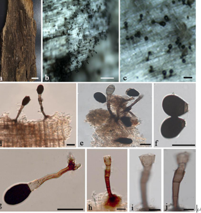
Pseudocercospora diospyri-japonicae (BPI 1109707 – holotype). A. Conidiophore fascicles. B. Conidiophores. C. Conidiophores, in the middle with attached young conidium. D. Conidia. Scale bar = 10 µm. U. Braun del.


