Diaporthe chongqingensis Y.S. Guo & G.P. Wang 2020
MycoBank MB830656
Holotype: China, Chongqing City, on branches of P. pyrifolia cv. Huanghua, 29 Mar. 2017, Y.S. Guo (holotype HMAS 248148, culture ex-type CGMCC 3.19603 = PSCG 435); ibid., culture PSCG436.
Morphological description
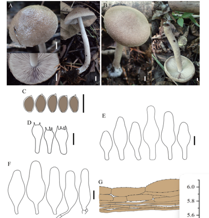
Sexual morph not observed. Asexual morph on alfalfa stems. Pycnidial conidiomata globose, solitary or aggregated, wrapped in hyphae embedded in alfalfa stems surface, grey to black, 285–744 μm diam, yellowish translucent conidial drops exuded from the ostioles. Conidiophores hyaline, smooth, 1-septate, densely aggregated, unbranched, ampulliform, 6.5–12.5 × 2–6 μm. Conidiogenous cells phialidic, hyaline, terminal, cylindrical, straight, 14–26 × 1.5–2.5 μm, tapered towards the apex. Alpha conidia hyaline, aseptate, fusiform, biguttulate or multi-guttulate, acutely round at one end, 5.5–7.5 × 2–3 μm, mean ± SD = 6.4 ± 0.5 × 2.3 ± 0.2 μm, L/W ratio = 2.8 (n = 50). Beta and gamma conidia not observed.
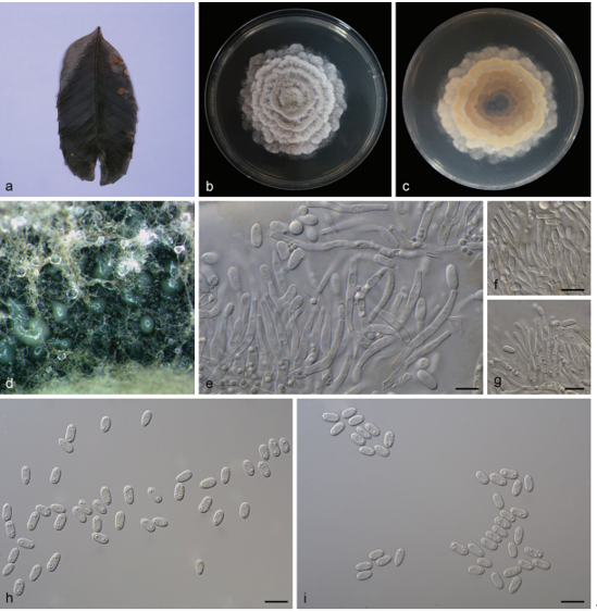
Culture characteristics — Colony on PDA with flattened mycelium, white, smoke grey in the centre, reverse with smoke grey coloured pigments formed in the shape of a concentric ring pattern. Colony diam 40–49 mm in 3 d at 28 °C. On OA, colony with entire margin, grey olivaceous in the centre and white margin, reverse grey olivaceous pigments formed in the centre.
Habitat: On branches of P. pyrifolia cv. Huanghua.
Distribution: In China.
GenBank Accession: ITS: MK626916; CAL: MK691209; HIS: MK726257; TEF: MK654866; TUB: MK691321
Notes: Diaporthe chongqingensis is introduced based on the multi-locus phylogenetic analysis, with two isolates clustering separately in a well-supported clade (BI/ML/MP = 1/100/100). Diaporthe chongqingensis is most closely related to D. fusicola, but distinguished based on ITS and TEF loci from D. fusicola (96.6 % in ITS and 97 % in CAL) by 24 nucleotides in the concatenated alignment, in which 15 are distinct in the ITS region, six in the TEF region and three in the TUB region. Morphologically, D. chongqingensis differs from D. fusicola in its smaller alpha conidia (5.5–7.5 × 2–3 vs 5.5–9 × 2–3 μm).
Reference: Y.S. Guo1,2,3,4, P.W. Crous 5,6,7,8, Q. Bai 4 et al.
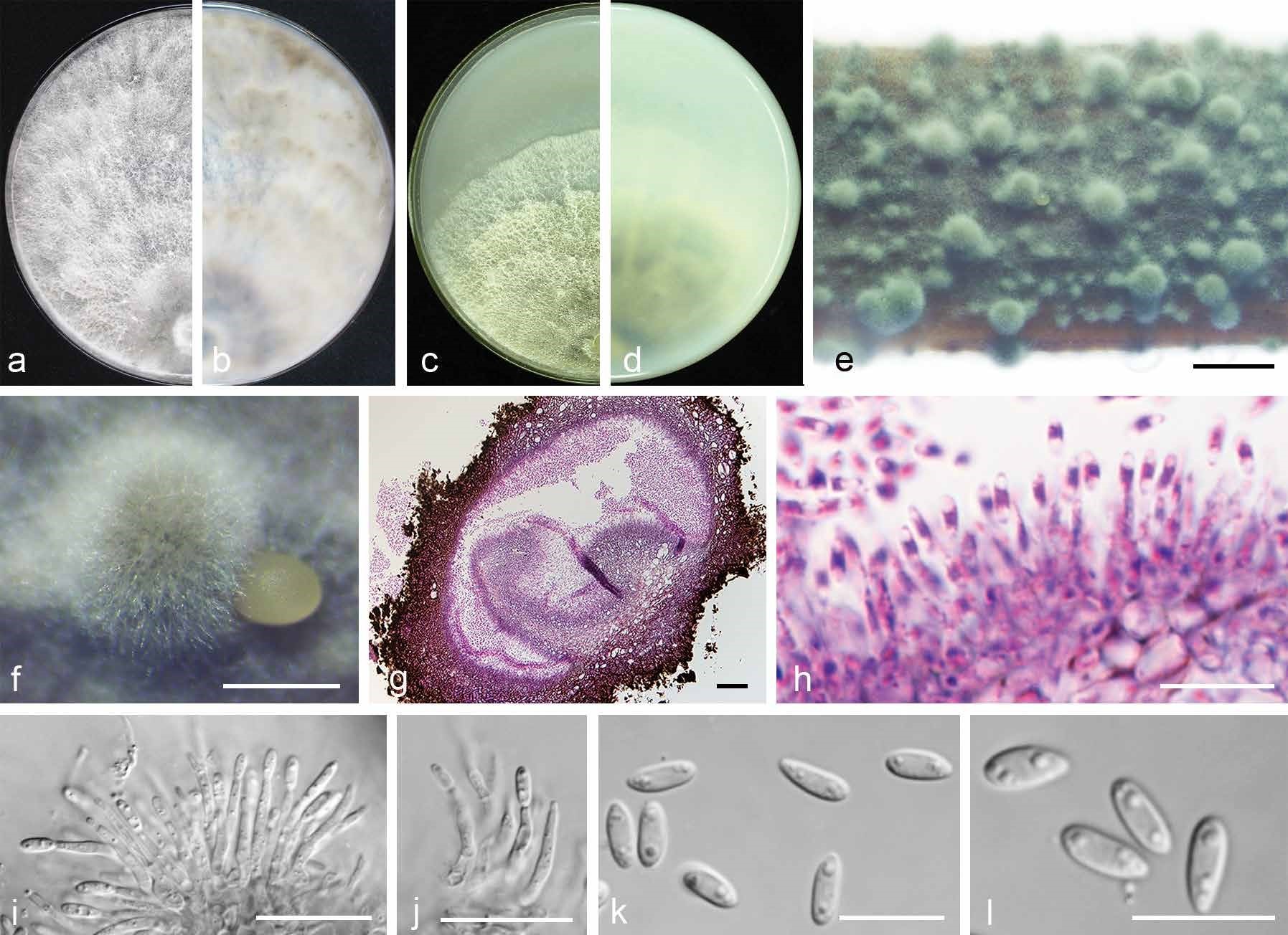
Diaporthe chongqingensis (CGMCC 3.19603). a–d. Front and back view, respectively of colonies on PDA (a, b) and OA (c, d); e. conidiomata on alfalfa stems; f. conidiomata; g. section view of conidiomata; h–j. conidiophores; k–l. alpha conidia. — Scale bars: e = 2 mm; f = 500 μm; g = 50 μm; i–j = 20 μm; h, k–l = 10 μm.