Diaporthe acuta Y.S. Guo & G.P. Wang 2020
MycoBank MB830655
Holotype: China, Hubei Province, Wuhan City, on branches of P. pyrifolia cv. Cuiguan, 1 Sept. 2014, Q. Bai (holotype HMAS 248147, culture ex-type CGMCC 3.19600 = PSCG 047); ibid., culture PSCG 045 and PSCG 046.
Morphological description
Sexual morph not observed. Asexual morph on alfalfa stems. Pycnidial conidiomata globose or irregular, solitary or aggregated, exposed on the alfalfa stems surface, dark brown to black, 230–544 μm diam. Alpha conidia hyaline, aseptate, fusiform to oval, acutely round at both ends, bi- or multi-guttulate, 6–9.5 × 2–3 μm, mean ± SD = 7.8 ± 0.6 × 2.6 ± 0.2 μm, L/W ratio = 3 (n = 50). Beta and gamma conidia not observed.
Culture characteristics — Colonies on PDA with flattened mycelium, aerial mycelium scarce, flocculent scattered distribution, surface and reverse luteous. Colony diam 63–67 mm in 3 d at 28 °C. On OA with aerial mycelium white, fluffy, sulphur yellow pigment accumulation in the centre, pure white at the colony margin.
Habitat: On branches of P. pyrifolia cv. Cuiguan.
Distribution: In China.
GenBank Accession: ITS: MK626957; CAL: MK691125; HIS: MK726161; TEF: MK654802; TUB: MK691225
Notes: Three isolates were identified as D. acuta in a wellsupported clade in the D. arecae species complex. This species is most closely related to D. pescicola, D. fulvicolor and D. spinosa, but easily distinguished from D. pescicola by 85 nucleotides difference in the concatenated alignment (40 in the ITS region, 6 TEF, 38 CAL and 1 TUB), from D. fulvicolor by 82 nucleotides difference (43 in the ITS region, 3 TEF, 17 CAL, 3 HIS and 16 TUB) and from D. spinosa by 24 nucleotides difference (13 in the ITS region, 7 CAL and 4 TUB). Moreover, D. acuta differs from D. pescicola in morphology, namely having smaller conidiomata (230–544 vs 637–881 μm), larger alpha conidia (6–9.5 × 2–3 vs 6–8 × 2–2.5 μm) (Table 4) and lacking beta conidia. However, its pycnidial conidiomata are larger than those of D. fulvicolor (230–544 vs 174–316 μm) and D. spinosa (230–544 vs 124–172 μm).
Reference: Y.S. Guo1,2,3,4, P.W. Crous 5,6,7,8, Q. Bai 4 et al.
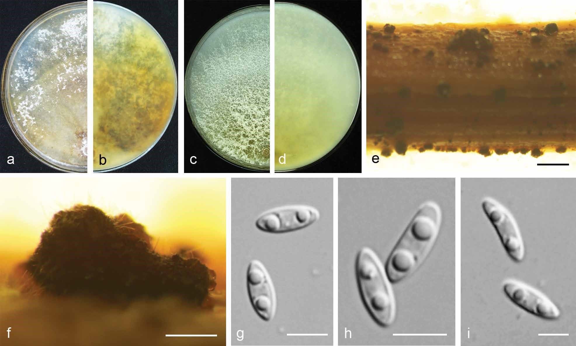
Diaporthe acuta (CGMCC 3.19600). a–d. Front and back view, respectively of colonies on PDA (a, b) and OA (c, d); e. conidiomata on alfalfa stems; f. conidiomata; g–i. alpha conidia. — Scale bars: e = 1 mm; f = 200 μm; g–i = 5 μm