Diaporthe fulvicolor Y.S. Guo & G.P. Wang 2020
MycoBank MB830657
Holotype: China, Hubei Province, Wuhan City, on branches of P. pyrifolia cv. Cuiguan, 1 Sept. 2014, Q. Bai (holotype HMAS 248149, culture ex-type CGMCC 3.19601 = PSCG 051); ibid., culture PSCG 057.
Morphological description
Sexual morph not observed. Asexual morph on alfalfa stems. Pycnidial conidiomata globose or irregular, solitary or aggregated, exposed on the alfalfa stems surface, dark brown to black, 174–316 μm diam. Conidiophores hyaline, smooth, 1-septate, densely aggregated, unbranched, cylindrical, straight, 5.5–8 × 2.5–3.5 μm. Conidiogenous cells phialidic, hyaline, terminal, ampulliform, 6.5–10 × 1.5–2.5 μm, tapered towards the apex. Alpha conidia hyaline, aseptate, fusiform to oval, acutely round at both ends, biguttulate or multi-guttulate, 7–9 × 2–3 μm, mean ± SD = 7.8 ± 0.4 × 2.5 ± 0.2 μm, L/W ratio = 3.1 (n = 50). Beta and gamma conidia not observed.
Culture characteristics — Colonies on PDA with aerial mycelium white, fluffy, reverse tawny pigment accumulation in the centre, surrounded by amber, pure white at the colony margin. Colony diam 52–55 mm in 3 d at 28 °C. On OA with entire margin, greyish yellow-green in the centre and white margin.
Habitat: On branches of P. pyrifolia cv. Cuiguan.
Distribution: In China.
GenBank Accession: ITS: MK626859; CAL: MK691132; HIS : MK726163;TEF: MK654806; TUB: MK691236
Notes: Diaporthe fulvicolor forms an independent clade in the D. arecae species complex (Fig. 4) and is phylogenetically distinct from D. pescicola and D. spinosa (described below). Diaporthe fulvicolor can be distinguished from D. pescicola in CAL and TUB loci by 57 nucleotide differences in concatenated alignment (40 in CAL and 17 in TUB), and from D. spinosa in CAL loci by 15 nucleotides (93 % in CAL). Moreover, D. fulvicolor differs from D. pescicola in having smaller conidiomata (174–316 vs 637–881 μm), and larger alpha conidia (7–9 × 2–3 vs 6–8 × 2–2.5 μm). Furthermore, D. fulvicolor differs from D. spinosa in its longer alpha conidia (7–9 × 2–3 vs 5.5–8 × 2–3.5 μm).
Reference: Y.S. Guo1,2,3,4, P.W. Crous 5,6,7,8, Q. Bai 4 et al.
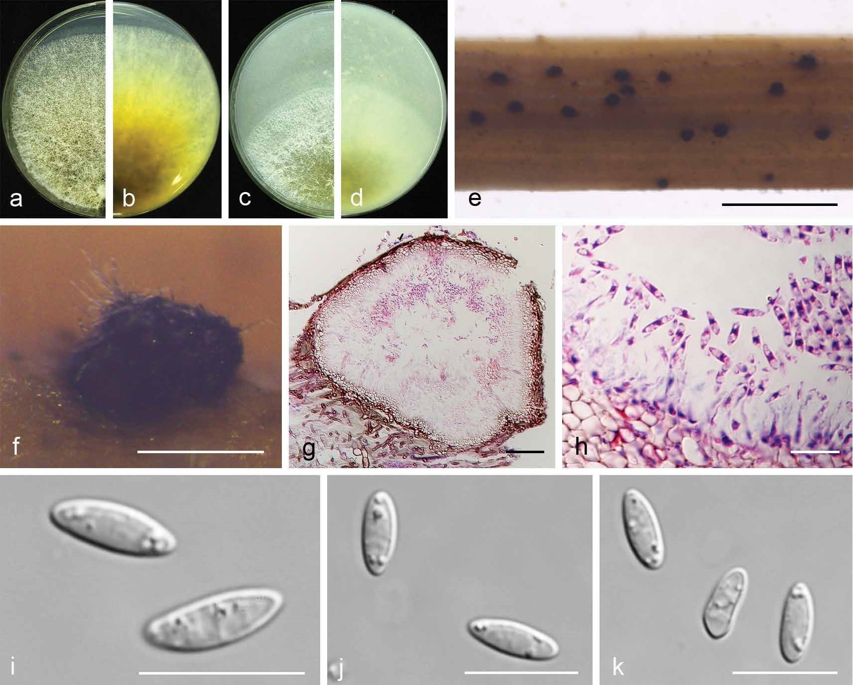
Diaporthe fulvicolor (CGMCC 3.19601). a–d. Front and back view, respectively of colonies on PDA (a, b) and OA (c, d); e. conidiomata on alfalfa stems; f. conidiomata; g. section view of conidiomata; h conidiophores; i–k. alpha conidia. — Scale bars: e = 2 mm; f = 200 μm; g = 50 μm; h–k = 10 μm.