Xenoastrosphaeriella trochus (D. Hawksw.) Phookamsak, H.B. Jiang, & K.D. Hyde, comb. nov. 2020
Index Fungorum number: IF 111138; Facesoffungi number: FoF 08164
Holotype: China, Yunnan Province, Xishuangbanna, Mengla County, Xishuangbanna tropical botanical garden, on dead stem of Thysanolaena maxima, 27 April 2017, R. Phookamsak, IS001 (KUN-HKAS 107533), living culture, KUMCC 18-0194.
Morphological description
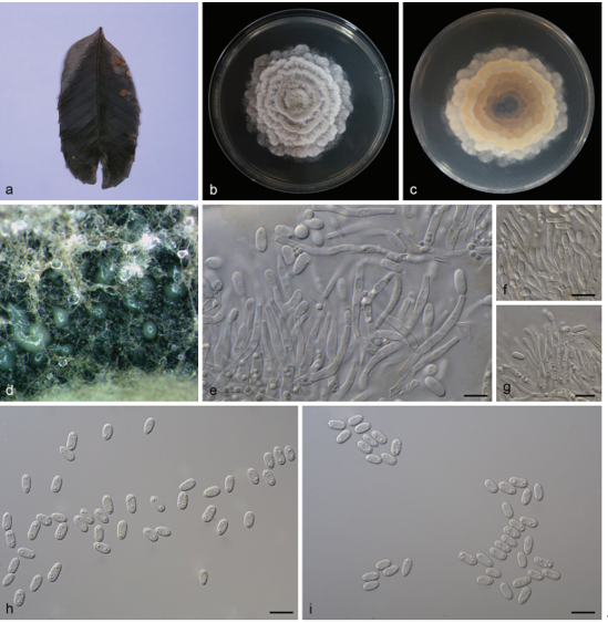
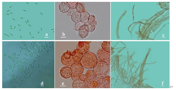
Saprobic on Thysanolaena maxima (Poaceae), visible as black, opaque, cone-like, on the host surface. Sexual morph: Ascomata 570–810 μm high, 590–760 μm diam., carbonaceous, dark brown to black, opaque, solitary to gregarious, erumpent through the outer layers of the host tissue, becoming superficial, easily broken, conical to mammiform, with host cortex persisting as ruptured, reflexed, stellate, host remnants, around the base, uni-loculate, rarely bi-loculate, glabrous, ostiolate, with a minute papilla. Peridium 40–110 μm wide, of unequal thickness, poorly developed at the base, thicker at sides towards the apex, composed of small, dark opaque, melanized cells of textura prismatica or palisade-like. Hamathecium comprising dense, 0.5–1.5 μm wide, filiform, trabeculate, anastomosing at the apex, pseudoparaphyses, embedded in a hyaline gelatinous matrix. Asci (125–)160–190(–215) × 10–12(–14) μm (x̅ = 172.5 × 11.9 μm, n = 30), 8-spored, bitunicate, fissitunicate, subcylindrical to cylindric-clavate, short pedicellate, apically rounded, with welldeveloped ocular chamber. Ascospores (40–)45–55(–57) × 4.5–6 μm (x̅ = 48.2 × 5.6 μm, n = 30), overlapping 1–2-seriate, narrowly elongate fusiform with acute ends, brown to reddish brown, paler at the end cells, (3–)5-septate, slightly constricted at the septa, smooth-walled, with conspicuous mucilaginous sheath surrounded ascospores. Asexual morph: Undetermined. Culture characteristics – Ascospores germinated on WA after 8 hours at 25°C under a dark condition. Colonies on PDA reaching 35–40 mm diam. after 4 weeks at 25–30°C, colonies circular, dense, slightly raised to convex, or dome-shaped, dull, surface slightly rough with small turfs and brown droplets, edge entire, velvety to floccose; colony from above, drak-green at the margin, grey-greenish at the center; from below, dark brown to black at the margin, paler at the center; not producing pigmentation in agar.
Habitat: on dead stem of Thysanolaena maxima
Distribution: Chile, China (Xishuangbanna, Yunnan), Colombia, Ecuador, French Guiana, Japan, Indonesia, South Africa, Taiwan, Uganda (Hawksworth 1981, Hawksworth & Boise 1985, Hyde & Fröhlich 1998).
GenBank Accession: LS MT659668;SSU MT659669;tef MT653597;rpb-2 MT653598
Notes: Astrosphaeriella trochus was designated for Melanomma trochus by Hawksworth (1981). However, the species should be transferred to Xenoastrosphaeriella based on multi-gene phylogeny and morphology. Xenoastrosphaeriella trochus is similar to X. tornata in having conical to mammiform, carbonaceous ascomata, cylindric-clavate asci and broadly fusiform, reddish brown ascospores, but differs in septa number and with or without a sheath (Phookamsak et al. 2015b, this study). Based on the present phylogeny (Fig. 53), X. tornata and X. trochus grouped together with high statistic support (100 % MLBS, 1.0 PP).
Reference: Hongsanan S, Hyde KD, Phookamsak R et al. 2020 – Refined families of Dothideomycetes: Dothideomycetidae and Pleosporomycetidae.

Xenoastrosphaeriella trochus (KUN-HKAS 107533). a Appearance of ascomata on host surface. b, c Section through ascoma. d, e Section through peridial structures. f Asci with trabeculate pseudoparaphyses embedded in a mucilaginous matrix. g, j Asci. h Ascus stained by congo red. i Ascus stained by India ink. k–o Ascospores. p Ascospore stained by India ink. q Germinated ascospore. r, s Culture characteristic on PDA after 4 weeks (r = from above, s = from below). Scale bars: b, c = 200 μm, e, f = 50 μm, d, g–j, q = 20 μm, k–p = 10 μm.