Sanguinoderma laceratum Y.F. Sun & B.K. Cui, sp. nov. 2020
MycoBank MB828436
Holotype: China, Yunnan Province, Baoshan, Gaoligongshan Nature Reserve, on fallen angiosperm trunk, 25 Sept. 2009, B.K. Cui, Cui 8155 (BJFC; isotype IFP).
Morphological description
Basidiomata annual, laterally stipitate, soft corky. Pileus multiple and superposed, flabelliform or reniform, up to 7.5 cm diam and 1 cm thick. Pileal surface dark cinnamon when dry, dull, tomentose, with faint concentric zones and radial wrinkles; margin acute, entire, wavy and incurved when dry. Pore surface pale buff to greyish brown when fresh, colour changing to blood red when bruised, becoming dark brown to nearly black; pores irregular, 2–3 per mm; dissepiments thin, lacerate when dry. Context straw colour to pale brown, with pale grey melanoid lines, soft corky, up to 4 mm thick. Tubes concolorous with context, woody hard, becoming fascicular when dry, up to 6 mm long. Stipe concolorous with pileal surface, cylindrical and hollow, up to 8.5 cm long and 4 mm diam. Hyphal system trimitic; generative hyphae with clamp connections, all hyphae IKI–, CB+; tissues darkening in KOH. Generative hyphae in context colourless, slightly thick-walled, 3–4 μm diam; skeletal hyphae in context pale yellow, thick-walled with a wide to narrow lumen or subsolid, arboriform branched and flexuous, 3–7 μm diam; binding hyphae in context pale yellow, subsolid, branched and flexuous, up to 2 μm diam. Generative hyphae in tubes colour- less, slightly thick-walled, 3–4 μm diam; skeletal hyphae in tubes pale yellow, thick-walled with a wide to narrow lumen or subsolid, arboriform branched and flexuous, 3–6 μm diam; binding hyphae in tubes pale yellow, subsolid, branched and flexuous, 1–2 μm diam. Pileal cover composed of clamped generative hyphae, thin- to thick-walled, apical cells clavate, inflated and flexuous, pale yellow to yellowish brown, about 25–40 × 6–13 μm, forming a regular palisade. Cystidia or cys tidioles absent. Hymenium collapsed in the studied sample, basidia and basidioles not seen. Basidiospores subglobose to broadly ellipsoid, pale yellow, IKI–, CB+, with double and slightly thick walls, exospore wall smooth, endospore wall with spinules, (10.2–)10.5–12.3(–12.5) × (8.6–)9–10.5(–11.1) μm, L = 11.38 μm, W = 9.71 μm, Q = 1.17 (n = 60/1). Under SEM, exospore wall semi-reticulate, badly worn, endospore wall with long and slightly thick coniform spinules tightly arranged.
Habitat: on fallen angiosperm trunk
Distribution: Yunnan Province, China.
GenBank Accession: ITS MK119851a; nLSU MK119928a
Notes: Sanguinoderma laceratum can be distinguished by the superposed pileus with slight concentric zones and lacerate pores with thin dissepiments. Sanguinoderma laceratum shares a deeply worn exospore wall with S. bataanense, but the latter has globose to subglobose basidiospores with distinctly thick walls with a verrucose exospore wall (Fig. 8a–b). In the phylogenetic analyses, S. laceratum was shown to be a distinct lineage in Sanguinoderma (Fig. 1, 2).
Reference: Y.-F. Sun1,2, D.H. Costa-Rezende3, J.-H. Xing1 et al.
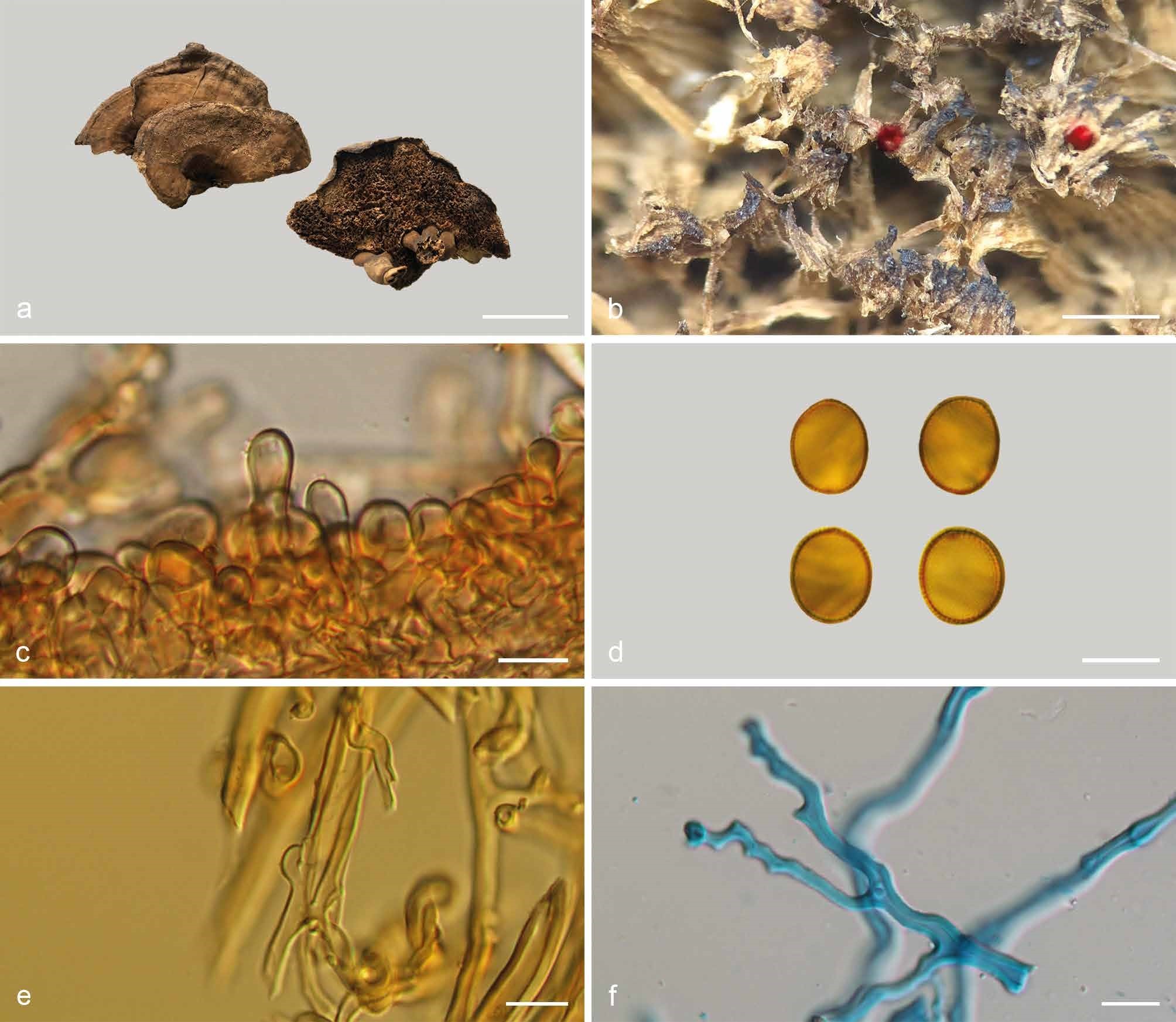
Basidiomata and microscopic structures of Sanguinoderma laceratum (Cui 8155). a. Basidiomata; b. pores; c. apical cells from pileal cover; d. basidiospores; e. generative hyphae from tubes; f. skeletal hyphae from context. — Scale bars: a = 3.5 cm; b = 1.5 mm; c–f = 1