Aspergillus phialosimplex Z.F. Zhang & L. Cai, sp. nov. 2020
Index Fungorum number: 556396; Facesoffungi number: FoF 08429
Holotype: CHINA, Sichuan, Huaying, Liujia Cave, N 30.41°, E 106.878°, on plant debris, May 2016, Z.F. May 2016, Z.F. Zhang, LC12658; Yunnan, Yuxi, Niumo Cave, N 28.192°, E 102.842°, on plant root, May 2016, Z.F. Zhang, LC12625.
Morphological description
Hyphae hyaline, septate, smooth, branched, 1.0–3.5 μm wide, sometimes swollen, up to 7.0 μm. Asexual morph Conidiogenous cells simple phialides arising laterally on vegetative hyphae. Phialides cylindrical, occasionally ampulliform, variable in length, smooth, hyaline, 2.5–8.5 µm long, 1.0–2.0 µm diam. Conidia formed in long chains, subglobose to globose, thick-walled, rough initially, then becoming smooth with age, hyaline, 3.5–5.5 µm ( x̄ ± SD = 4.7 ± 0.42, n = 60). Sexual morph not observed. Culture characteristics—Colonies on PDA attaining 20–29 mm diam. after 4 weeks, flat, felty to pulverulent, margin slightly undulate, brown (7C5) to dark brown (7F7) from margin to center. Reverse pale brown (6B3) to dark brown (7F8). Colonies on OA attaining 20–28 mm diam. after 4 weeks, flat, margin entire, white to pale lavender (6B2), aerial mycelia sparse. Reverse white to pale brown. Colonies on SNA attaining 42–46 mm diam. after 4 weeks, flat, pulverulent, margin unclear, white. Reverse white. Sporulation within 3 weeks.
Habitat: on plant debris.
Distribution: Sichuan, Huaying, Liujia Cave, China.
GenBank Accession:
Notes: Aspergillus phialosimplex is phylogenetically allied to A. phialiformis (Fig. 9), but they can be easily distinguished (see notes of A. phialiformis).
Reference: Zhi‑Feng Zhang ,Shi‑Yue Zhou ,Lily Eurwilaichitret al.
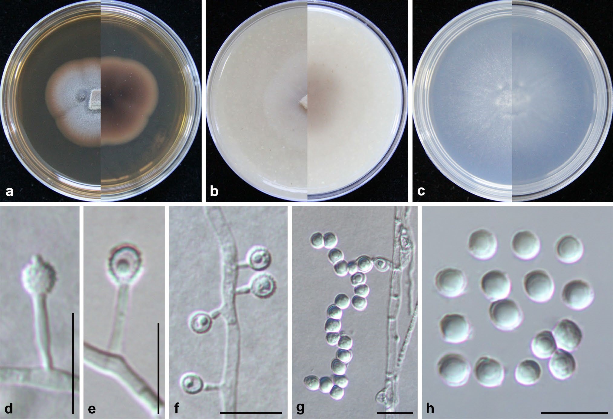
Aspergillus phialosimplex (from ex-holotype CGMCC3.19637). a–c upper and reverse views of cultures on PDA, OA and SNA 4 weeks after inoculation; d–g phialides and conidia; h. conidia. Scale bars: d–h 10 µm