Acrogenospora obovoidspora D.F. Bao, Z.L. Luo, K.D. Hyde & H.Y. Su, sp. nov. 2020
Index Fungorum number: IF 557602; Facesoffungi number: FoF 04691
Holotype: CHINA, Yunnan Province, Dali, Huadianba Mountain, saprobic on decaying wood submerged in a stream, 9 December 2017, Z.L. Luo, S-1614 (MFLU 20–0295, holotype); ex-type culture, MFLUCC 18–1622.
Morphological description
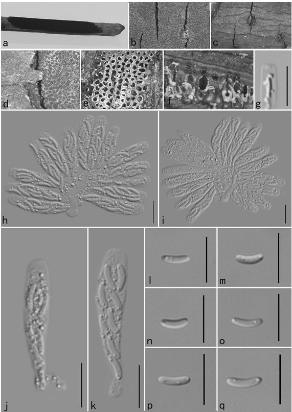
Saprobic on submerged decaying wood. Sexual morph: Undetermined. Asexual morph: Colonies effuse on natural substrate, hairy, dark brown. Mycelium partly immersed, partly superficial, composed of septate, brown to dark brown, branched, smooth hyphae. Conidiophores 209–277 × 7.5–10 µm (x¯ = 243 × 8.8 µm, n = 15), mononematous, macronematous, solitary, erect, straight or slightly flexuous, cylindrical, unbranched, brown to dark brown, paler toward apex, septate, smooth. Conidiogenous cells holoblastic, monoblastic, integrated, initially terminal, later becoming intercalary, cylindrical, smooth, pale brown, proliferating percurrently. Conidia 32.5–37.5 × 27– 32 µm (x¯ = 35 × 29.6 µm, n = 30) wide, acrogenous, solitary, oval to broadly ellipsoidal, base truncate, aseptate, olivaceous brown to black, thick-walled, smooth.
Habitat: on decaying wood submerged in a stream.
Distribution: Inner Mongolia Autonomous Region, China.
GenBank Accession: LSU MT340736; SSU MT340747; RPB2 MT367163; TEF-1α MT367155
Notes: Acrogenospora obovoidispora is similar to A. gigantospora in having mononematous, macronematous, conidiophores and solitary, aseptate conidia. However, Acrogenospora obovoidspora differs from A. gigantospora in having solitary conidiophores and oval to broadly ellipsoidal, olivaceous brown to black conidia, while conidiophores of A. gigantospora are single or in groups of 2–4, and conidia are broadly obovoid to subspherical, dark brown to black (Ma et al., 2012).
Reference: Dan-Feng Ba1,2,3, Eric H. C. McKenzie4, D. Jayarama Bhat5 et al.
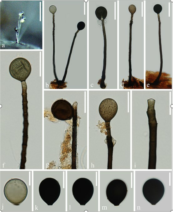
Acrogenospora obovoidispora. (MFLU 20–0295, holotype) (a) Colony on wood. (b-e) Conidiophores with conidia. (f-i) Conidiogenous cells with conidia. (j-n) Conidia. Scale bars. (a) 200µm. (b-e)50µm. (f-h)30µm. (i-n)20µm.