Exophiala nagquensis W. Sun, L. Su, M.C. Xiang and X.Z. Liu, sp. nov. 2020
MycoBank MB 824211
Holotype: China: Tibet: Nagqu, 30◦190 N, 90◦400 E, 4591 m a.s.l., from rock, 4 August 2013, Meichun Xiang, (HMAS 245344 (dried culture)–holotype and CGMCC 3.17333–ex-type culture).
Morphological description
Hyphae poorly branched, smooth-walled, septate, light olivaceous brown,2.0–3.1-µm-diam. Budding cells consisting of subspherical cells occasionally observed (Figure 22I,H). Conidiogenous cells arising from undifferentiated hyphae, terminal or intercalary, brown, cylindrical, poorly differentiated, sometimes with sympodial conidiogenesis (Figure 22C–F). Conidia 0-1-septate, hyaline to pyrite yellow, broadly ellipsoid, obovoid, and global 4.8–10.4 × 2.6–5.0 µm (x = 7.8 × 3.3 µm, n = 20) (Figure 22G,J).Culture characters: Colony on MEA growing slowly, attaining 34-mm-diam. after four weeks at 25 ◦C. Colony surface velvety with olivaceous grey short aerial hyphae and margin irregular, reverse olivaceous black (Figure 22A,B). No diffusible pigment produced. Minimum 4 ◦C, optimum at 20–25 ◦C, and maximum 28 ◦C.
Habitat: from rock
Distribution: China
GenBank Accession:
Notes: E. nagquensis forms a sister clade to E. brunnea (CBS 587.66), with 96% ML and 1.00 Bayesian support (Figure 5). However, they can be distinguished by conidial morphology, and E. brunnea has narrower conidia (2–3 µm vs. 2.6–5.0 µm) compared with the new species. In the meanwhile, the budding cells are absent for E. brunnea [36] but present for the new species.
Reference: Wei Sun , Lei Su , Shun Yang et al.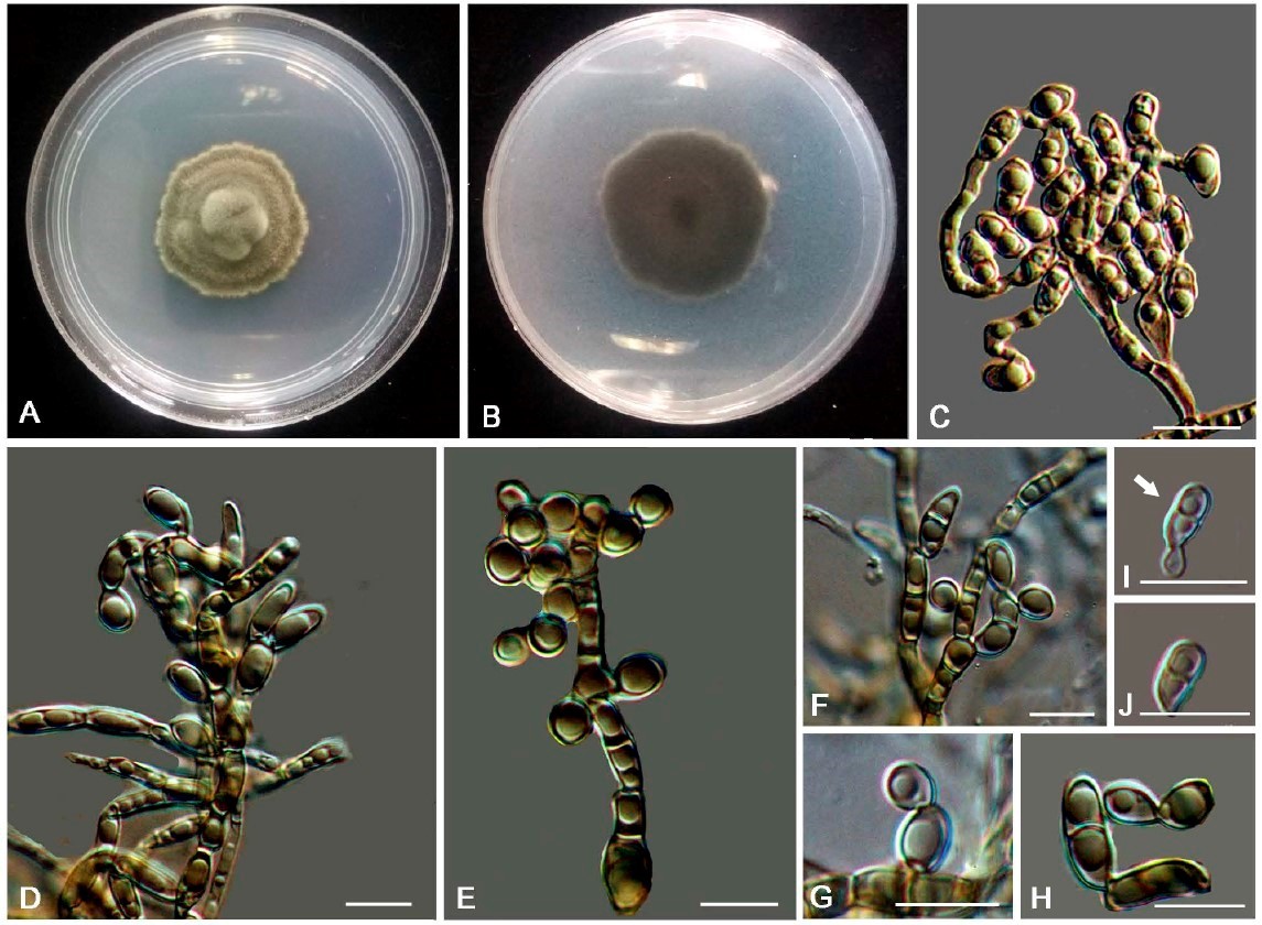
Exophiala nagquensis (CGMCC 3.17333). (A,B) Forward and reverse of colony on MEA.
(C–F) Conidiophores with conidiogenous cells. (G–J) Conidia and budding cells. Scale bars: (C–J) = 10 µm.