Knufia calcarecola L. Su, W. Sun and M.C. Xiang, sp. nov. 2020
MycoBank MB 810892
Holotype: China: Beijing City: Beijing Botanical Garden, dry climate with fog and haze weather in autumn and winter, 39◦590 N, 116◦120 E, 89 m a.s.l., ornamental rocks, from limestone, 12 April 2011, Lei Su, (HMAS 245385 (dried culture)–holotype and CGMCC 3.17218–ex-type culture).
Morphological description
Hyphae catenulate, becoming moniliform with age, septate, constricted at the septa, 3.5–7.1-µm (x = 5.3 µm, n = 20)-wide, thick-walled, pale brown to greyish brown, guttulate, enteroblastically proliferating (Figure 10F). Pycnidia initials arising from a few irregular cells formed by repeated division of a few hyphae cells, 90.2–186.8-µm-wide, 127.2–232.9-µm-high (x = 140.2 × 200.6 µm, n = 10), sub-globose to globose, brown to dark–brown (Figure 10C,D). Conidiogenous cells hyaline to pale brown, cylindrical to sub-globose, 6.9–8.2 × 12.3–23.2 µm (x = 8.4 × 16.5 µm, n = 10) (Figure 10E). Conidia gemmating proliferation is commonly present laterally along the hyphae (Figure 10H). Conidia hyaline, usually aseptate, occasionally 1-septate, constricted at the septa, smooth-walled, cylindrical to clavate, 4.8–6.2 × 8.5–14.1 µm (x = 5.3 × 10.8 µm, n = 20)(Figure 10I). Terminal multicellular bodies ellipsoidal to muriform, brown to dark brown, 7.3–21.4 × 10.4–22.6 µm (x = 18.5 × 19.4 µm, n = 10) (Figure 10G).Culture characteristics: Colonies on MEA growing slowly, attaining 15-mm-diam. after 20 weeks at 25 ◦C, compact, raised centrally, greyish brown, aerial mycelia sparse to scant, black in reverse (Figure 10A,B). Minimum 4 ◦C, optimum at 20–25 ◦C, and maximum 28 ◦C.
Habitat: from limestone
Distribution: Beijing (China).
GenBank Accession:
Notes: K. calcarecola is phylogenetically close to K. petricola. However, pycnidia were only observed in K. calcarecola.
Reference: Wei Sun , Lei Su , Shun Yang et al.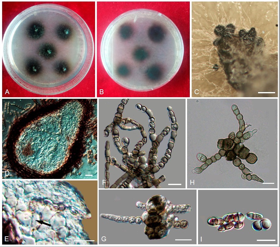
Knufia calcarecola(CGMCC3.17218). (A,B)Colonyforwardandreverseafter20weeksonMEA. (C) Pycnidia with hyaline surface hyphae. (D) Vertical section through pycnidium. (E) Conidiogenous cells (arrow). (F) Catenated, moniliform hyphae. (G) Multicellular bodies. (H) Conidia germination. (I) Conidia. Scale bars: (C) = 50 µm, (D) = 5 µm, and (E–I) = 10 µm.