Exophiala cinerea W. Sun, M.C. Xiang and X.Z. Liu, sp. nov. 2020
MycoBank MB 824205
Holotype: China: Guizhou Province: Leishan County, Leigongshan National Nature Reserve, 26◦230 N, 108◦120 E, 2152 m a.s.l., from rock, 13 October 2014, Meichun Xiang, (HMAS 247722 (dried culture)–holotype and CGMCC 3.18778–ex-type culture).
Morphological description
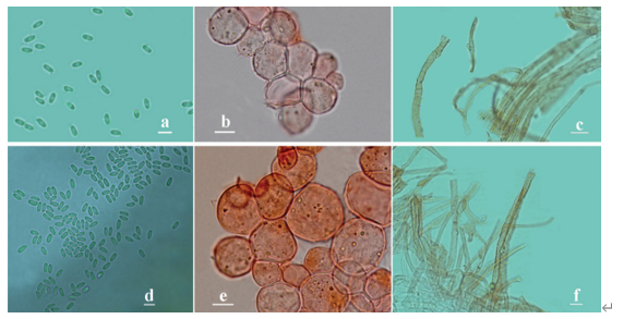
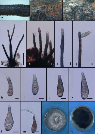
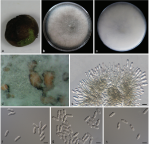
Hyphae poorly branched, spirally twisted hyphae present, smooth and thin-walled, septate, guttulate, hyaline to light olive-grey, 1.0–4.1-µm-diam. (Figure 19B,C). Yeast cells absent. Conidiogenous cells, ellipsoidal-shaped, ovoidal to elongate, intercalary or terminal (Figure 19E). Conidia, one-celled, surface smooth, thin-walled, arising alongside the hyphae or at the apex. Solitary, cylindrical, or truncate at the base, 3.6–8.8 × 2.7–5.4 µm (x = 5.6 × 3.7 µm, n = 20) (Figure 19D,F–H).Culture characters: Colony on MEA growing slowly, attaining 15-mm-diam. after four weeks at 25 ◦C. Colony surface olivaceous grey to black, raised centrally, flat and glossy near the periphery, velvety with grayish-brown short aerial hyphae and irregularly lobate margin; reverse grey black (Figure 19A). No diffusible pigment produced. Minimum 4 ◦C, optimum at 20–25 ◦C, and maximum 28 ◦C.
Habitat: from rock
Distribution: China
GenBank Accession:
Notes: E. cinerea forms a sister clade of E. ellipsoidea with 99% ML and 1.00 Bayesian support (Figure 5). E. cinerea is morphologically similar to E. ellipsoidea. However, E. cinerea has longer conidia (3.6–8.8 × 2.7–5.4 µm vs. 2.1–6.4 × 1.1–4.0 µm) than that of E. ellipsoidea.
Reference: Wei Sun , Lei Su , Shun Yang et al.
Exophiala cinerea (CGMCC 3.18778). (A) Forward (left) and reverse (right) of colony on MEA. (B,C) Spirally twisted hyphae. (D) Conidia alongside hyphae. (E) Conidial apparatus with conidia.
(F–H) Conidia. Scale bars: (B–H) = 10 µm.