Exophiala clavispora W. Sun, M.C. Xiang and X.Z. Liu, sp. nov.
2020
MycoBank MB 824207
Holotype: China: Tibet: Nyingchi, Qinghai-Tibet Plateau, 20◦450 N, 94◦450 E, 3448 m a.s.l., from rock, 2 August 2013, Meichun Xiang, (HMAS245389 (dried culture)–holotype and CGMCC3.17517–ex-type culture).
Morphological description
Hyphae composed by cylindrical to elongate cells, hyaline, guttulate, septate, 1.1–3.2-µm-wide, spirally twisted hyphae frequently observed (Figure 20C). Yeast cells absent. Conidiogenous cells ovoidal to elongate, olivaceous grey, 6.2–10.1-µm-long (Figure 20E). Conidia arising alongside the hyphae or at the apex, cylindrical to clavate, truncate at the base, 5.9–11.9 × 1.8–3.7 µm (x = 8.1 × 2.3 µm, n = 20) (Figure 20D,E).Culture characters: Colony on MEA growing slowly, attaining 12-mm-diam. after four weeks at 25 ◦C. Colony surface cerebriform, arise centrally, velvety with grayish short aerial hyphae and lobate margin; reverse deep olivaceous gray (Figure 20A,B). Mycelium submerged into the agar. No diffusible pigment produced. Minimum 4 ◦C, optimum at 20–25 ◦C, and maximum 28 ◦C.
Habitat: from rock
Distribution: China
GenBank Accession:
Notes:
Reference: Wei Sun , Lei Su , Shun Yang et al.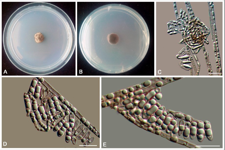
Exophiala clavispora (CGMCC 3.17517). (A,B) Forward and reverse of colony on MEA.
(C) Spirally twisted conidial chains. (D,E) Conidia alongside hyphae. Scale bars: (C–E) = 10 µm.