Microdochium indocalami S.T. Huang, J.W. Xia, X.G. Zhang, W.X. Sun & Z. Li, sp. nov. 2020
MycoBank No: 835766
Holotype: China, Yunnan Province: Xishuangbanna Tropical Botanical Garden, Chinese Academy of Sciences, on diseased leaves of Indocalamus longiauritus. 16 April 2019, S.T. Huang, HSAUP1016, holotype, ex-type living culture SAUCC1016.
Morphological description
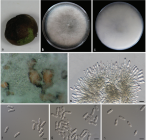
Colonies on PDA attaining 46.1–51.2 mm in diameter after 7 days, formed a conspicuous concentric circle, periphery of aerial mycelium cottony, centre with scarce aerial mycelium, white initially, then becoming greyish sepia after 25 days. Some aerial hyphae aggregated and form a sporodochium within 15 days or longer. Mycelium composed of hyaline, immersed and superficial, smooth, branched, septate, 2.0–3.0 μm wide hyphae. Due to the soluble pigment secreted, reverse white to salmon. Conidiophores straight or slightly curved, aseptate, aggregated in the aerial mycelium, often reduced to conidiogenous cells borne directly from the hyphae. Conidiogenous cells terminal or intercalary, mono-or polyblastic, denticulate, smooth, hyaline, cylindrical, straight or bent, 11.0–28.3 × 1.5–2.9 μm. Conidia cylindrical, clavate to obovoid, 1–3-septate, 13.0–15.5 × 3.5–5.5 μm, base usually flattened 0.5– 1.0 μm. Sometimes borne directly from the mycelial hyphae. Sexual morph: unknown.Culture characteristics. Colonies on OA 62.0–64.0 mm in diameter after 7 days, centre with aerial mycelium cottony, periphery with scarce aerial mycelium. Mycelium mostly immersed, hyphae hyaline, septate, smooth, exudate and soluble pigment produced, reverse white initially, then becoming pale mouse-grey in periphery and mousegrey in center. Sporodochia formed on agar surface. Colonies on MEA 50.8–52.7 mm in diameter after 7 days, aerial mycelium abundant, with concentric rings, white to pale pink, periphery with cottony aerial mycelium, centre with scarce aerial mycelium, exudate absent. Reverse white to pale pink with age.
Habitat: on diseased leaves of Indocalamus longiauritus.
Distribution: China
GenBank Accession: LSU MT199878; ITS MT199884; TUB2 MT435653; RPB2 MT510550
Notes: Microdochium and allied genera were revised by Hernández-Restrepo et al. (2016). Phylogenetic analysis of a combined four gene showed that M. indocalami (strain SAUCC1016) formed a separated branch as the clade of M. fisheri, M. lycopodinum, M. phragmites, and M. rhopalostylidis with good support (ML-BS: 100% and BYPP: 1.00) (Fig. 1). Additionally, conidiogenous cells of M. indocalami are terminal or intercalary, denticulate, cylindrical which are similar to the species in this clade. The size of conidia and the number of septa of conidia reported for M. fisheri (7.0–12.0 × 3.0– 4.0 μm, 0–1-septate), M. lycopodinum (8.0–15.5 × 2.5–4.0 μm, 0–1-septate), M. phragmites (10.0–14.5 × 2.0–3.0 μm, 0–1-septate), and M. rhopalostylidis ((13.0–) 16.0–20.0 (–23.0) × (2.5–) 3.0 (–4.0) μm, 1–3-septate) (Hernández-Restrepo et al. 2016; Crous et al. 2019) were different to M. indocalami (13.0–15.5 × 3.5–5.5 μm, 1–3-septate).
Reference: Huang S, Xia J, Zhang X et al. (2020) Two new species of Microdochium from Indocalamus longiauritus in south-western China.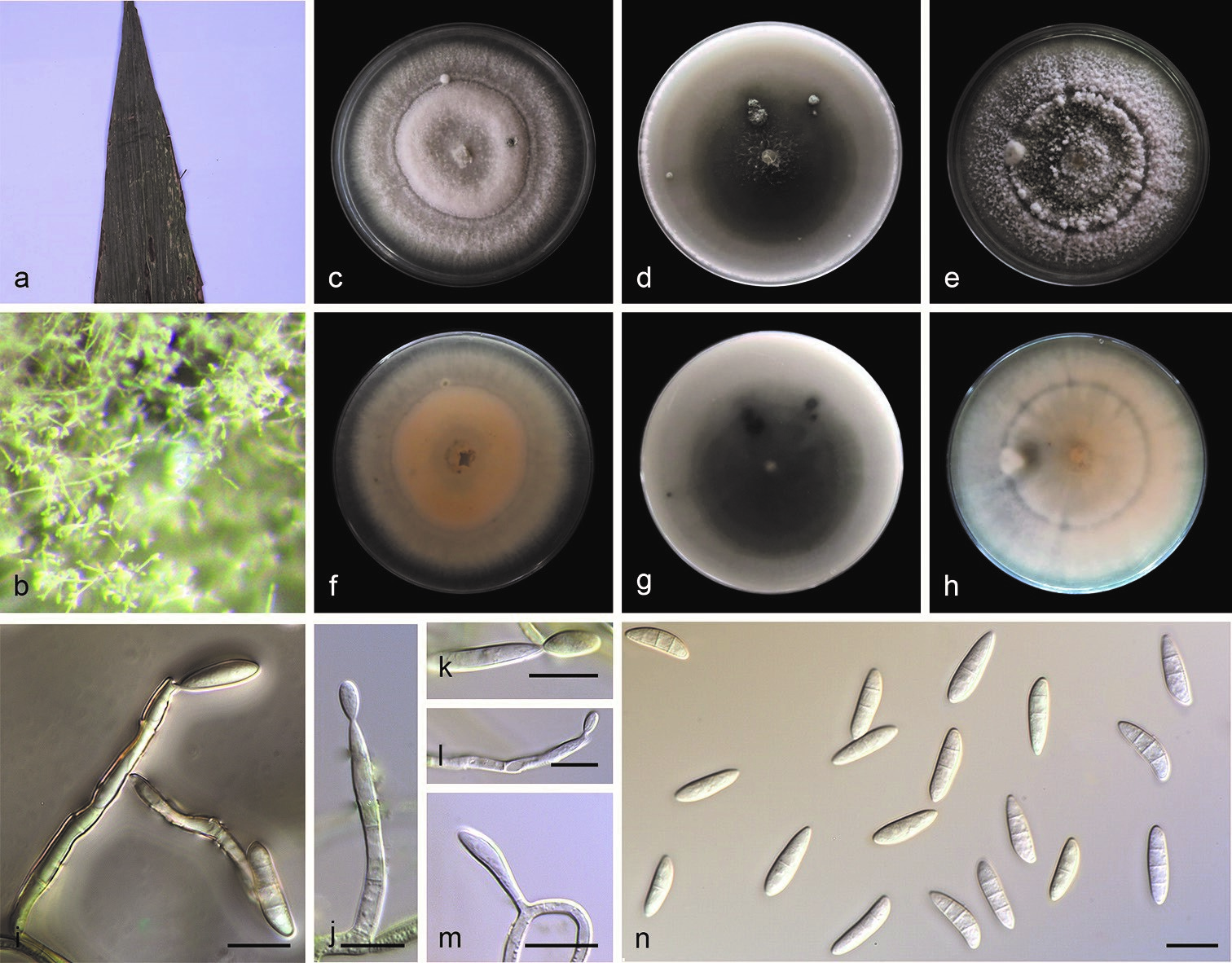
Microdochium indocalami (SAUCC1016) a leaves of host plant b colony overview c–e surface of colony after 15 days on PDA (c), OA (d), MEA (e) f–h reverse of colony after 15 days on PDA (f) OA (g) MEA (h) i–m conidiophores and conidiogenous cells n conidia. Scale bars: 10μm (i–n).