Microdochium sp. indet. 2020
MycoBank
Holotype: China, Yunnan Province: Xishuangbanna Tropical Botanical Garden, Chinese Academy of Sciences, on diseased leaves of Indocalamus longiauritus. 16 April 2019, S.T. Huang, HSAUP1017, living culture SAUCC1017.
Morphological description
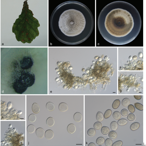
Colonies on PDA attaining 73.9–80.4 mm in diameter after 15 days, felty to cottony, flat, margin entire or dentate, white, aerial mycelium abundant. Mycelium superficial, hyphae hyaline, septate, branched, smooth-walled. Reverse white to pale yellow, with yellow pigment produced with aging. Aerial hyphae aggregated to form numerous chlamydospores on agar surface. Chlamydospores thick-walled, terminal or intercalary, more frequently arranged in chains than clusters. Conidiophores not observed. Colonies on OA attaining 79.9–81.7 mm in diameter after 15 days, fluffy, margin entire, white. Reverse white. Colonies on MEA attaining 73.2–78.4 mm in diameter after 15 days, flat, with pale pink inconspicuous concentric circle near the centre, margin entire and white, aerial mycelium abundant.
Habitat: on diseased leaves of Indocalamus longiauritus.
Distribution: China
GenBank Accession: LSU MT199879; ITS MT199885; TUB2 MT435654
Notes: Strain SAUCC1017 failed to produce conidia and lacks a complete morphological description. It formed a conspicuous independent lineage from other Microdochium species in the tree. ITS sequence BLASTn search of SAUCC1017 showed many different species with 97% identity. BLASTn searches with LSU (GenBank MH869857) sequences result in 99% identity with M. bolleyi (CBS 172.63) and TUB2 (GenBank AB625368) sequences result in 99% identity with Xylaria cubensis (strain BCC 18758). Thus, here we listed it as an unidentified species.
Reference: Huang S, Xia J, Zhang X et al. (2020) Two new species of Microdochium from Indocalamus longiauritus in south-western China.
Microdochium sp. (SAUCC1017) a leaves of host plant b colony overview c–e surface of colony after 15 days on PDA (c) OA (d) MEA (e) f–h reverse of colony after 15 days on PDA (f) OA (g) MEA (h) i–k chlamydospores. Scale bars: 10μm (i–k).