 109
109
Russula luofuensis B. Chen & J. F. Liang, sp. nov.2021
MycoBank No: MB838836
Holotype: China. Guangdong Province, Huizhou City, Boluo County, Luofu Moun tain Provincial Nature Reserve, 23°15'47.13"N, 114°3'45.42"E, 90 m asl., in mixed Fagace ae forests of Cyclobalanopsis and Castanopsis, 22 August 2020, leg. CB446 (RITF4708).
Morphological description
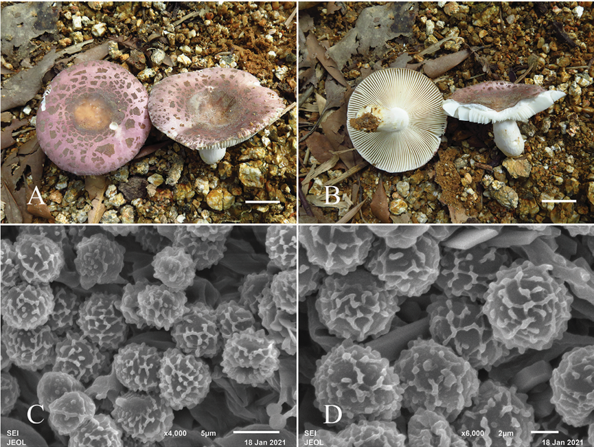
Sexual morph: Basidiomata medium-sized to large; pileus 35–80 mm in diameter; initially hemispheric when young, applanate to convex, convex with a depressed center after mature; margin incurved, not cracked, striation short and inconspicuous; surface dry, glabrous, peeling to 1/2 of the radius, cracking and broken into small golden brown patches, patches crowded towards the center, with smaller patches towards the margin; grayish yellow to brownish orange in the center, purplish gray to grayish magenta towards the margin. Lamellae adnate to subfree, 2–4 mm deep, 8–10 at 1 cm near the pileus margin, white to cream; lamellulae absent; furcations occasional near the stipe; edge entire and concolor. Stipe 30–50 × 10–25 mm, cylindrical, slightly inflated towards the base, white, with yellow ish tinge at the base, and medulla initially stuffed becoming hollow. Context 2–3 mm thick in half of the pileus radius, white, unchanging when bruised, taste mild, odor inconspicuous. Spore print pale yellowish.
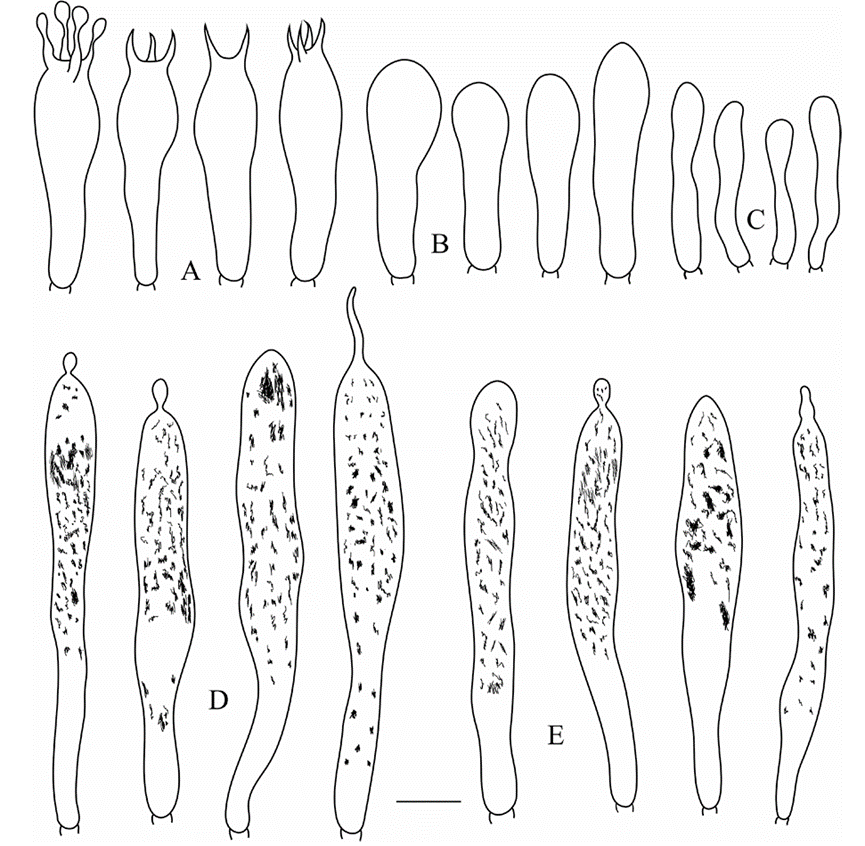
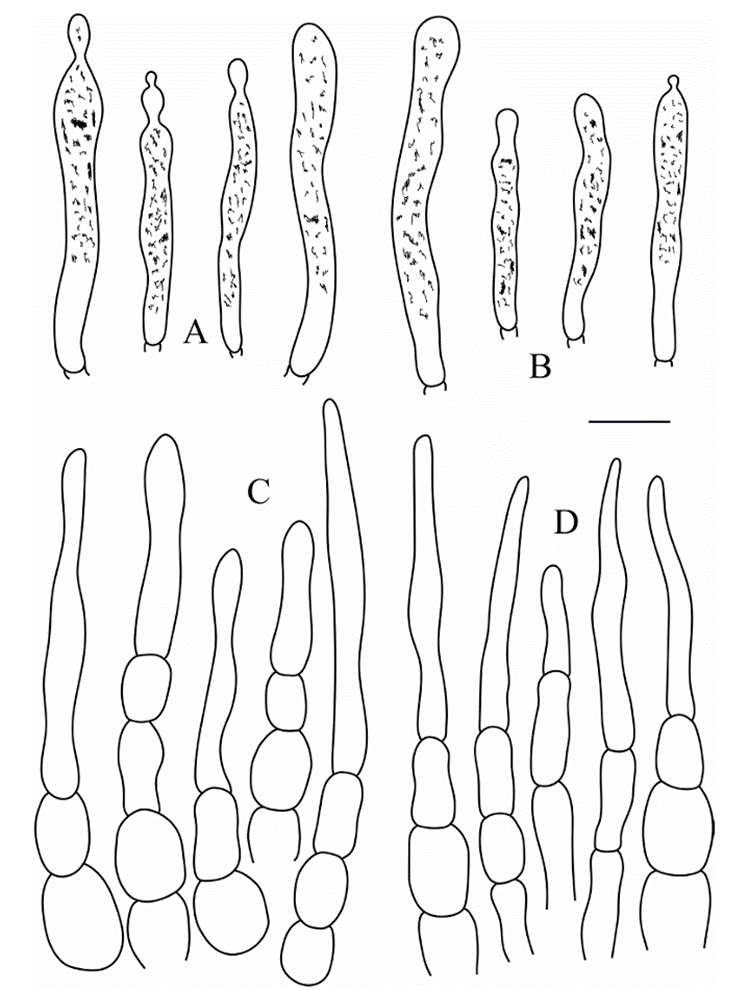
Basidiospores (5.0–)5.8–6.6–7.5(–8.6) × (4.5–)5.4–6.2–7.0(–8.0) μm, Q = (1.0–)1.02–1.08–1.14(–1.26), subglobose to broadly ellipsoid; ornamentation of me dium-sized, moderately distant to dense [6–8(–9) in a 3 μm diameter circle] amyloid warts or spines, 0.3–0.6 μm high, locally reticulate, frequently fused in short or long chains [2–3(–4) in the circle], occasionally to frequently connected by line connections [1–2(–3) in the circle]; suprahilar spot medium-sized, amyloid. Basidia (35.0–)36.7 39.8–42.8(–45.5) × (9.0–)9.5–10.0–10.5(–11.2) μm, mostly 4-spored, some 2- and 3-spored, clavate; basidiola clavate or subcylindrical, ca. 6.5–11.5 μm wide. Hymenial gloeocystidia on lamellae sides dispersed to moderately numerous, ca. 600–900/mm2, (59.0)63.2–71.3–79.3(83.6) × (7.0)7.7–8.8–9.9(10.5) μm, clavate or narrowly clavate, apically mainly obtuse, occasionally acute, often with 3–10 μm long appendage, thin walled; contents heteromorphous or granulose, mainly in the middle and upper part, turning reddish black in SV. Hymenial gloeocystidia on lamellae edges often smaller, (49.5–)56.2–64.3–72.4(–80.2) × (6.2–)7.3–8.3–9.4(–10.0) μm, clavate, or subcylindri cal, sometimes fusiform, apically mainly obtuse, occasionally mucronate, sometimes with 3–6 μm long appendage thin-walled; contents heteromorphous, turning reddish black in SV. Marginal cells (15.2–)19.8–23.5–27.2(–30.6) × (3.5–)4.0–4.8–5.6(–7.0) μm, subcylindrical or clavate, often flexuous. Pileipellis orthochromatic in cresyl blue, not sharply delimited from the underlying context, 260–300 μm deep, two-layered; su prapellis 120–150 μm deep, hyphal endings composed of inflated, ellipsoid or globose cells with attenuated terminal cells; subpellis 120–160 μm deep, composed of repent, intricate, 2–6 μm wide hyphae. Hyphal terminations near the pileus margin typically unbranched, occasionally flexuous, thin-walled; terminal cells (9.2–)18.6–28.2–37.8( 50.8) × (3.2–)3.9–5.0–6.1(–8.2) μm, mainly narrowly lageniform, occasionally clavate or cylindrical, apically attenuated or constricted, occasionally obtuse; subterminal cells frequently shorter and wider, ca. 4–9 μm wide, typically unbranched. Hyphal termina tions near the pileus center similar to those near the pileus margin; terminal cells (10.2 )18.4–27.4–36.4(–44.8) × (3.2–)3.6–4.7–5.8(–6.8) μm, mainly lageniform, occasion ally fusiform or subcylindrical, apically attenuated or constricted; subterminal cells often shorter and wider, rarely branched, ca. 4–7 μm wide. Pileocystidia near the pileus mar gin always one-celled, (23.3–)27.9–35.0–42.2(–47.5) ×3.5–4.8–6.0(–8.3) μm, mainly clavate, occasionally subcylindrical or fusiform, apically typically obtuse, occasionally acute, often with round or ellipsoid, 3–6 μm long appendage, thin-walled; contents heteromorphous or granulose, turning reddish black in SV. Pileocystidia near the pileus center similar in size, always one-celled, (24.6–)27.2–34.8–42.5(–48.2) × 3.0–4.2–5.4( 6.8) μm, thin-walled, mainly clavate, occasionally fusiform, apically often obtuse or oc casionally acute, occasionally with 2–4 μm long appendage, contents heteromorphous or granulose, turning reddish black in SV. Cystidioid hyphae In subpellis and context with heteromorphous contents, oleiferous hyphae in subpellis with refringent contents.
Asexual morph: Undetermined
Culture characteristics:
Habitat:
Distribution: China
GenBank Accession:
ITS MW646973; nrLSU MW646985; mtSSU MW646996; TEF1 MW650842
ITS MW646974; nrLSU MW646986; mtSSU MW646997; TEF1 MW650843
ITS MW646975; nrLSU MW646987; mtSSU MW646998; TEF1 MW650844
ITS MW646976; nrLSU MW646988; mtSSU MW646999; TEF1 MW650845
ITS MW646977; nrLSU MW646989; mtSSU MW647000; TEF1 MW650846
Notes: The combination of morphological features and phylogenetic analysis place R. luofuensis in subsect. Virescentinae. Phylogenetically, our new species R. luofuensis is clustered with R. albidogrisea with 89% bootstrap support and 1.00 posterior probabil ities, which is also from Guangdong Province of China. However, R. albidogrisea differs from R. luofuensis in having a white to grayish pileus with acute, even to slightly un dulate margin, often smaller basidiospores [(5.1–)5.3–5.6–6.0(–6.4) × (4.6–)4.8–5.1 5.3(–5.6) μm], longer hymenial gloeocystidia on lamellae sides (35–50 × 5–11 μm) and hymenial gloeocystidia on lamellae edges (37–46 × 9–12 μm, Das et al. 2017).
Given cracking surface, R. viridirubrolimbata, R. parvovirescens, R. virescens and R. crustosa Peck of subsect. Virescentinae resemble R. luofuensis. However, R. viridiru brolimbata, originally described from China, can be distinguished by a light yellowish olive to yellowish olive pileus center with a pinkish red to light jasper red margin and absence of hymenial gloeocystidia on lamellae edges (Ying 1983; Deng et al. 2020). T he American species R. parvovirescens possesses a greenish brown to metallic bluish green pileus with green patches (Buyck et al. 2006). Russula virescens (originally re ported from Europe) is distinct in its green to yellowish green pileus (Sarnari 1998). Russula albidogrisea, originally reported from North America, has a brownish-yellow, greenish or subolivaceous pileus with small spot-like areolae or pseudo-verrucae, short er basidia [(29–)30–32–33.5(–35) × (7.5–)8–9.5–10.5(–11) μm] and absence of hy menial gloeocystidia on the lamellar edges (Adamčík et al. 2018).
Reference: [1] Chen, B. , Song, J. , Chen, Y. , Zhang, J. , & Liang, J. . (2021). Morphological and phylogenetic evidence for two new species ofrussulasubg.heterophyllidiafrom guangdong province of china. MycoKeys, 82, 139-157.
Figure 2. Fruiting bodies (A, B) and basidiospores (C, D) of Russula luofuensis (RITF4708). Scale bars: 20 mm (A、B)
Figure 3. Russula luofuensis (RITF 4708) A basidia B basidiola C marginal cells D hymenial gloeocyst idia on lamellae sides E hymenial gloeocystidia on lamellae edges. Scale bar: 10 μm. 
Figure 4. Russula luofuensis (RITF 4708) A pileocystidia near the pileus margin B pileocystidia near the pileus center C hyphal terminations near the pileus margin D hyphal terminations near the pileus center. Scale bar: 10 μm.

