 103
103
Hirsutella kuankuoshuiensis X. Zou, J.J. Qu & Z.Q. Liang, sp. nov. 2021
MycoBank No: 819591
Holotype: China, Guizhou Province, Suiyang County, Kuankuoshui Nature Reserve (28°08'N, 107°02'E, approximately 1400 m a.s.l.), July 2012, collected by X. Zou. The holotype has been deposited at KIB (HKAS112885). Sequences from isolated strains (GZUIFR-2012KKS3-1, GZUIFR-2012KKS3-2 and GZUIFR-2012KKS3-3) have been deposited in GenBank.
Morphological description
Sexual morph: Undetermined
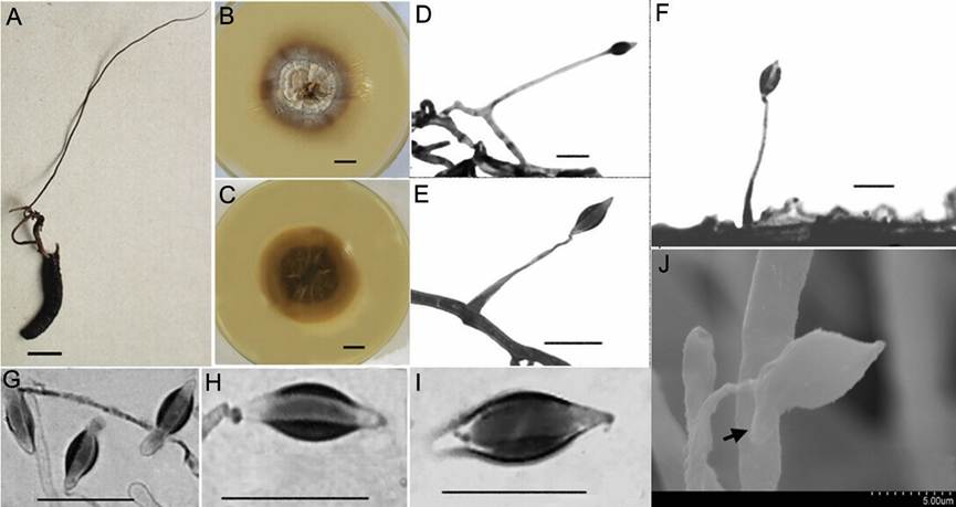
Asexual morph: Synnemata are single, extending from the head of insect; 8.6 cm long, dark brown and changing to brown towards the apex; no conidiation was ob served (Fig. 3A). The fungus spreads slowly on PDA agar at 20–22 °C and grows to a diam. of 22–30 mm after 14 d; the colony is round, centre of surface with brown dense bulges and grey-white sparse flocculent aerial hyphae. Colony margin is flat with radial groove; a large amount of brown pigment secreted into the medium causes the back of colony to appear dark brown; thickness 10–12 mm (Fig. 3B, C). Myce lium hyaline, smooth, septate, 1.5–3.0 μm wide. Conidiogenous cells monophialidic, hyaline, borne perpendicular or at an acute angle to the subtending hyphae. Phialides subulate or slender columnar, tapering gradually to a long and narrow neck, 30–45 × 1–3 μm long. Conidia clavate, narrow fusiform or botuliform without a diaphragm, 9.9–12.6 × 2.7–4.5 μm, single- or double-enveloped in a hyaline mucus, thickness 2.0–3.0 μm (Fig. 3D–J).
Culture characteristics:
Habitat: On the decaying leaves of broadleaved forests.
Distribution: Guizhou Province, China
GenBank Accession:
ITS KY415575; LSU KY415582; SSU -; RPB1 KY945360; TEF1α KY415590
ITS MF623038; LSU MF623044; SSU -; RPB1 -; TEF1α MF623048
ITS MF623039; LSU MF623045; SSU -; RPB1 -; TEF1α MF623049
1 Lepidoptera: GZUIFR 2012KKS3-2 Lepidoptera: GZUIFR 2012KKS3-3
Remarks:This species possesses two types of conidiogenous cells and long fusi form or clavate without diaphragm conidia (9.9–12.6 × 2.7–4.5 μm), which is ex tremely rare in Hirsutella species. In addition, H. kuankuoshuiensis could produce long thin synnemata on the culture media that contain few or no conidia.
Reference: [1] Qu, J. , Zou, X. , Cao, W. , Xu, Z. , & Liang, Z. . (2021). Two new species of hirsutella (ophiocordycipitaceae, sordariomycetes) that are parasitic on lepidopteran insects from china. MycoKeys, 82, 81-96.
Figure 3. Morphological characteristics of Hirsutella kuankuoshuiensis A the insect specimens with single and thin synnemata (HKAS112885) B, C colonial morphology on PDA agar media for 20 d B shows the front of the colony and C shows the back of the colony D–G LM images showing conidiogenous cells and conidia D, E the structure of conidiogenous cells on mycelia F the images of conidiogenous cells on synnemata (optical microscope) H–J conidial morphology (LM) G conidia with mucilage (SEM). Scale bars: 10 mm (A–C); bar of G was shown in the figure; the rest of the bars were 10 μm. LM, light micros copy; PDA, potato dextrose agar; SEM, scanning electron microscopy. Description. Synnemata are single, extending from the head

