 82
82
Tricholosporum guangxiense T.Bau et G.F.Mou, sp. nov.2021
Mycobank No: MB841851.
Holotype: China, Guangxi province, Chongzuo city, Ningming country, Nongang National Nature Reserve, 22°14′31.54″ N,107°03′50.14″ E,elevation167m,22August2021,Guang-fu MouHMJAU59028(Holotype HMJAU!).
Morphological description
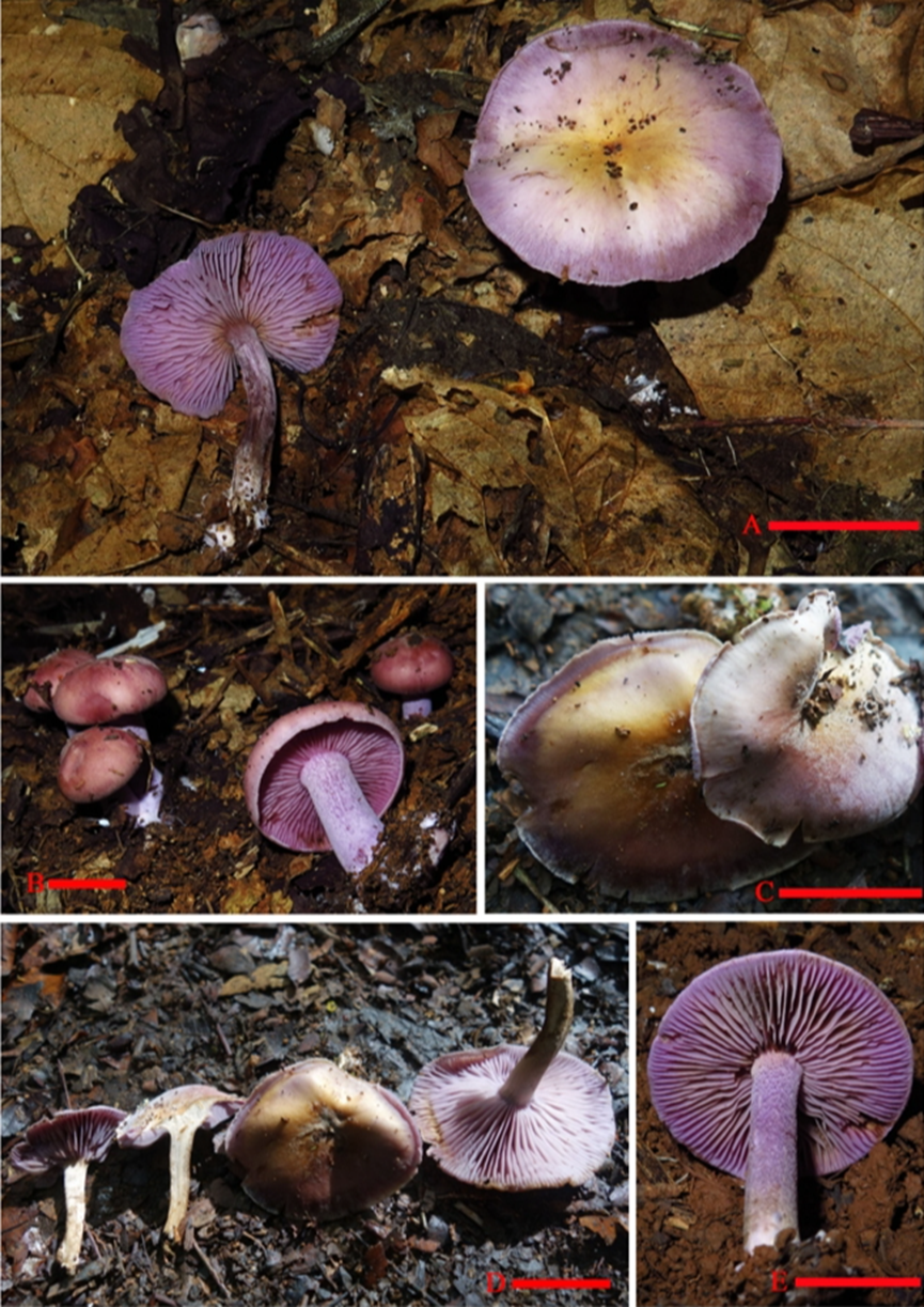
Sexual morph: Basidiomata tricholomatoid, solitary to gregarious (Figure 8). Pileus 43–55 mmindiam., convex to flat-hemispherical, becoming plane to depressed with age, margin smooth or with light short stripes, entire, incurved at first then straight to slightly reflexed, surface at first fibrillose-felted (due to very thin, white hairs), nonviscid; greyish ruby when young, light lilac to greyish violet near margin and central becoming light orange to brownish yellow with age. Context up to 4 mmthick, firm, whitish. Lamellae emarginate, with small decurrent tooth, close, 5 mminbroad, with 2–3 tiers of lamellulae; margins smooth; light violet to violet, turning orange to brownish orangewhen damaged. Stipe 30 to 50 mm long×5–8 mm in diam., stout, central, equal, dry, light violet to violet, covered by violet white to pale violet pruina, bruising dull. Basal with white rhizomorphs. Odor not distinct. Taste not recorded. Spore-print white.
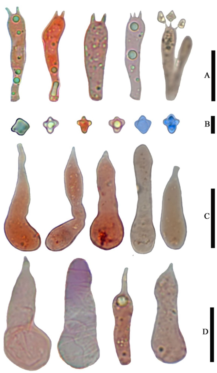
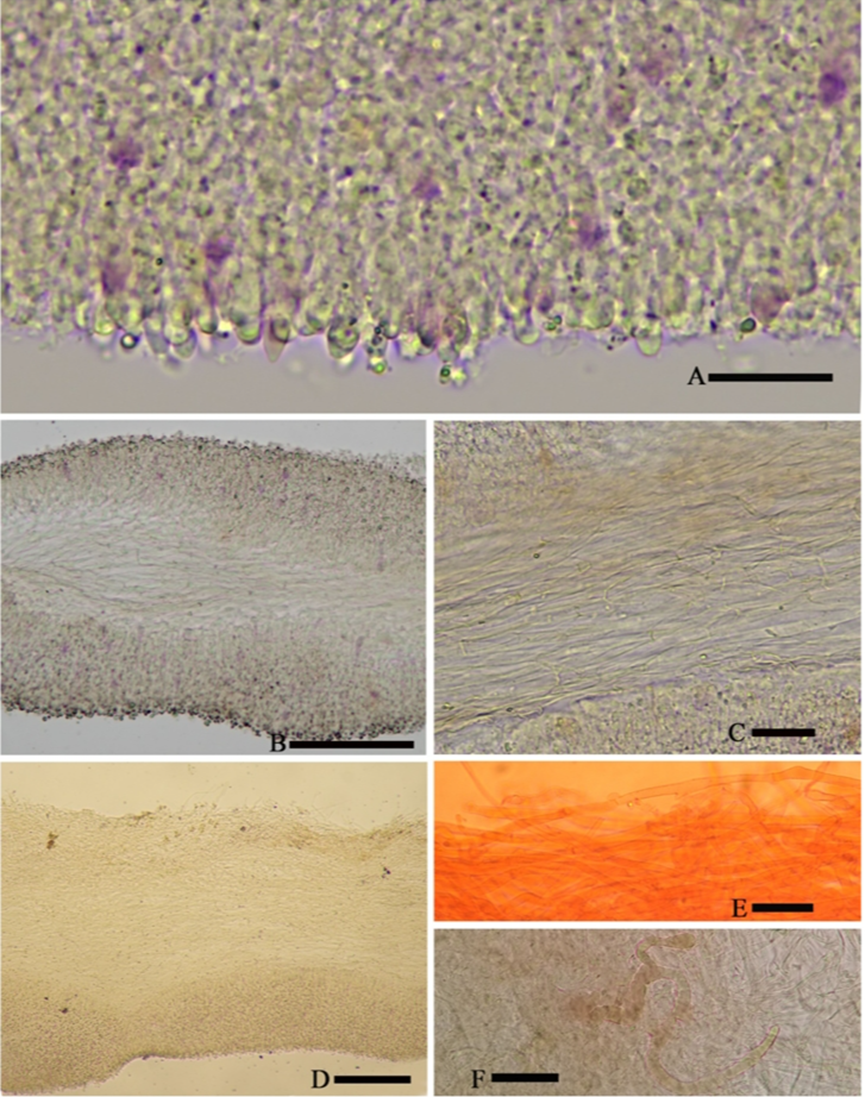
Basidiospores (4.0) 5.0–6.0 (7.0)×(3.6) 4.0–5.0 (5.4)μm, 5.4×4.7μm on average (Q =1.0–1.4, Qav = 1.2) [38/5/5], cruciform, hyaline, colorless, inamyloid, cyanophilous (Figure 9B 5–6), thin-walled, usually with a single large oil drop (Figure 9B). Basidia (21) 23–28 (32)×5–7μm [48/3/3], cylindric to narrowly clavate, thin-walled, color less, usually with one to multiple oil drops, two or four sterigmate (Figure 9A). Cheilo cystidia (23) 27–36 (40)×6–13 (14)μm, ampullaceous or clavate, with a swollen base and a neck, acute, mucronate or obtuse at apex, thin-walled, sometimes with purplish pink to greyish magenta pigment (Figures 9D and 10A). Pleurocystidia (35) 40–50 (60)×(8) 9–13 (14)μm, ampullaceous or clavate, with a swollen base and a neck, acute, mucronate or obtuse at apex, sometimes curved, thin-walled, sometimes with grey-violet pigment (Figure 9C). Hymenophoral trama 148–243μm broad, regular, hypha thin-walled (Figure 10B,C). Pileipellis an undifferentiated cutis, hyphae 4.6–5.8μm in diam., colorless (Figure 10D,E). Laticifers present, pale yellow, thick-walled, branched, 5–10μm in diam. (Figure 10F). Clamp connections present (Figure 10E).
Asexual morph: Undetermined
Culture characteristics:
Habitat: Scattered or gregarious on broad-leaved forest soil of karst areas; the associ ated tree species are Streblus tonkinensis, Wendlandia uvariifolia, Sterculia monosperma, Musa balbisiana, and Heptapleurum sp.
Distribution: So far, only known in Guangxi (China).
GenBank Accession:
Notes: Angelini et al., according to the size of the basidiospores, divided the species of Tricholosporum into two groups: group 1, the large-spored species with spores over 7μm in length, and group 2, species with small spores, usually under 6 m in length. According to the presence or absence of hymenial cystidia and whether grey-violet pigmentation was shown, they further divided group 2. Our specimens should obviously be categorized into group 2, subgroup 2.3: species with pigmented hymenial cystidia, which are grey-violet or brownish. Three species, T. palmense, T. violaceum, and T. caraibicum, were placed in this subgroup.
Our specimens differ from T. violaceum by their smaller and dry pileus (4.3–5.5 vs. 8–13 cm), thicker context (4 vs. 10–20 mm), narrower lamellae (5 vs. 15 mm), shorter stipe (5 vs. 5–11 cm), and larger spores (5.4×4.7 vs. 4.5×3.5μm, on average) [4]; differ from T. palmense by the unfurcate cystidia; differ from T. caraibicum by the pileus lacking color spots, central with obvious yellowish-brownish color when mature, the larger spores (5.4×4.7 vs. 4.1×3.8 μm,onaverage), and being cyanophilous [4].

Figure 8. Basidiomata of Tricholosporum guangxiense. Scale bar (A–D) = 2.5 cm. A from HMJAU59028 (Holotype HMJAU), (B) from HMJAU59027, (C,D) from HMJAU59023, and (E) from M2021082208 (IBK). Photos by Guang-fu Mou.
Figure 9. Microscopic features of Tricholosporum guangxiense, from HMJAU59028 (Holotype). (A–D) stained with 1% Congo Red solution. (B) (from left to right) 1–2 in pure water, 3–4 stained with 1% Congo Red solution, 5–6 stained by Cotton blue. (A) Basidia, (B) Basidiospores, (C) Pleurocystidia, and (D) Cheilocystidia. Scale bar (A) = 15μm, (B) = 5μm, (C,D) = 20μm. Photos by Guang-fu Mou.
Figure 10. Microscopic features of Tricholosporum guangxiense, from HMJAU59028 (Holotype). (A–D) F in pure water, (E) stained with 1% Congo Red solution. (A) Margin of lamella, (B,C) Hymenophoral trama, (D,E) Pileipellis and Hyphae of Pileipellis, and (F) Laticifer. Scale bar (A) = 15μm, (B) = 5μm, and (C–F) = 20μm. Photos by Guang-fu Mou.

