Arthrinium setariae C.M. Tian & N. Jiang, sp. nov 2021
MycoBank MB835609
Holotype: Dead culms of Setaria viridis in China.
Morphological description
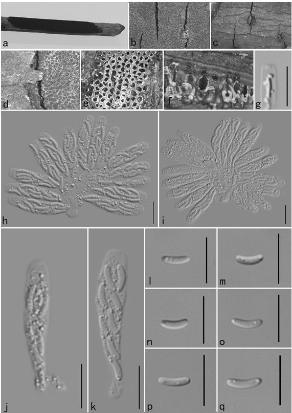
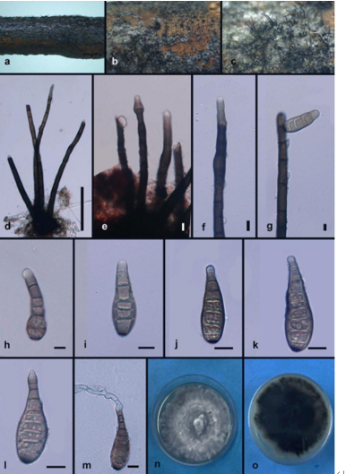
Stromata 350–1350 × 250–550 μm (n = 20), scattered to gregarious, partially immersed, becoming erumpent to superficial, raised, dark brown, in linear rows, with a slit-like opening, multi-loculate. Ascomata (80–)120–180(–200) μm (n = 30) diam., arranged in rows, clustered, gregarious, with 4–12 perithecia forming groups immersed in stromata to erumpent through host surface, ellipsoidal to subglobose, dark brown, membranous. Peridium 7–15 μm thick, consisting of 4–5 layers of cells arranged in textura angularis, externally dark brown, hyaline in the inner part. Ostiole raised from the centre of the ascomata, internally lined with periphyses, with a periphysate channel. Paraphyses intermingled among asci, not very prominent, hyphae[1]like, hyaline, smooth, septate, sparingly branched, thin-walled. Asci 60–95 × 15–21 μm (n = 20), 8-spored, unitunicate, clavate, broadly cylindrical, with an inconspicuous pedicel, rounded apex, thin-walled, without an apical apparatus. Ascospores (15–)18–23(–27) × (6–)8–11(–13) μm ( x = 21 × 9.5 μm, n = 50), 1-septate, ellipsoidal, with a smaller lower cell and a larger upper cell, hyaline, smooth-walled, with a faint gelatinous sheath surround the overall spore. Sporulating on PDA after 30 d. Hyphae 1.5–5 μm diam., hyaline, branched, septate. Conidiophores erect, hyaline, smooth. Conidiogenous cells 8–55 × 1–3.5 μm (n = 20), erect, hyaline to pale brown, smooth. Conidia 7.5–10.5 μm diam. ( x = 9.3 μm, n = 50), globose to subglobose, oval or irregular, brown, guttulate, usually with a longitudinal, hyaline, germ-slit.Cultures: Colonies fast growing on PDA, reaching 60 mm in 7 days at 25 °C, circular, convex, cottony, produce abundant red pigments and color the media to red. Sporulation after 30 d at 25 °C.
Habitat: Dead culms of Setaria viridis.
Distribution: CHINA, Beijing, Beijing Forestry University.
GenBank Accession: ITS MT492004; tub2 MT497466; tef1 MW118456.
Notes: Arthrinium setariae was the first species discovered from Setaria viridis (Poaceae). However, host information was not valid to separate Arthrinium species. From the phylogram (Fig. 1), A. jiangxiense collected from Maesa (Myrsinaceae) was the most related species. Arthrinium setariae differs from A. jiangxiense in having larger conidia (7.5–10.5 μm diam in A. setariae vs. 4.5–7 μm diam in A. jiangxiense) (Wang et al. 2018).
Reference: Jiang, N. , & Tian, C. M. . (2021). The holomorph of arthrinium setariae sp. nov. (apiosporaceae, xylariales) from china. Phytotaxa(483-), 483.
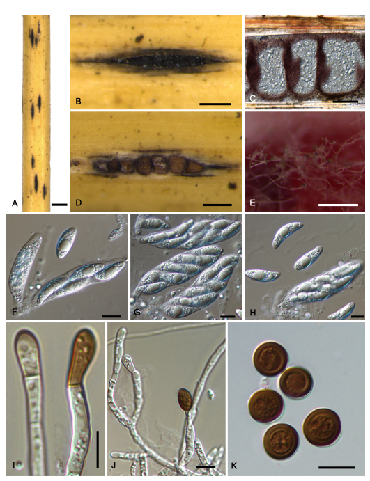 Arthrinium setariae. Morphology of Arthrinium setariae on culms of Setaria viridis and its conidial morph from PDA. A, B. Stromata on host; C, D. Transverse section of the stromata; E. Conidial morph on PDA; F–H. Asci and ascospores; I, J. Conidiogenous cells giving rise to conidia. K. Conidia. Bars: A = 500 μm; B–E = 50 μm; F–K = 10 μm.