Coryneum gigasporum C.M. Tian, Voglmayr & N. Jiang 2020
MycoBank MB824594.
Holotype: CHINA. SHAANXI PROVINCE: Shangluo City, chestnut plantation, 33°38′21.03″N, 109° 08′45.22″E, 2602 m asl, on branches of Castanea mollissima, N. Jiang, 8 Jul 2017 (holotype BJFC-S1425). Ex-type culture: CFCC 52319.
Morphological description
Sexual morph: Not observed.
Asexual morph: Conidiomata acervular, 0.8–1.5 mm wide, 0.5–1.0 mm high (x = 1.0 × 0.7 mm, n = 20), solitary, erumpent through outer periderm layers of host, scattered, surface tissues above slightly domed. Conidiophores 50–90 μm long, 3–6 μm wide (x = 55 × 5 μm, n = 20), unbranched, cylindrical, septate, hyaline at apex, pale brown at base. Conidiogenous cells holoblastic, integrated, indeterminate, cylindrical, expanding toward apices, hyaline to pale brown, smooth, with 0–1 percurrent extensions. Conidia (88–)93–108(–117) × (18–)19–21(–23) μm, L/W = (4.2–)4.6–5.4(–5.6) (n = 50), slightly curved or not, clavate, dark brown, smooth-walled, 7–9-distoseptate, apical cell with a hyaline tip, truncate and black at base.
Culture characters: On PDA at 25 C, colonies growing slowly and symmetrically, reaching 70 mm diam within 30 d, gradually becoming brownish gray in color with scant cottony aerial mycelium, asexual morphs developed after 2 mo.
Habitat: On branches of Castanea mollissima.
Distribution: In China.
GenBank Accession: ITS: MH683557; 28S: MH683565; TEF1α: MH685737; RPB2: MH685729
Notes: Conidial size and shape are a main character for species distinction in Coryneum (Sutton 1975). Coryneum gigasporum is unique for its large conidial size (88–117 × 18–23 μm) within the genus. The two other Coryneum species with very long conidia, C. megaspermum var. cylindricum from Quercus and C. stromatoideum from Tsuga canadensis, differ from C. gigasporum by longer and narrower conidia (TABLE 1).
Reference: Ning Jiang a, Hermann Voglmayr b, and Chengming Tian a
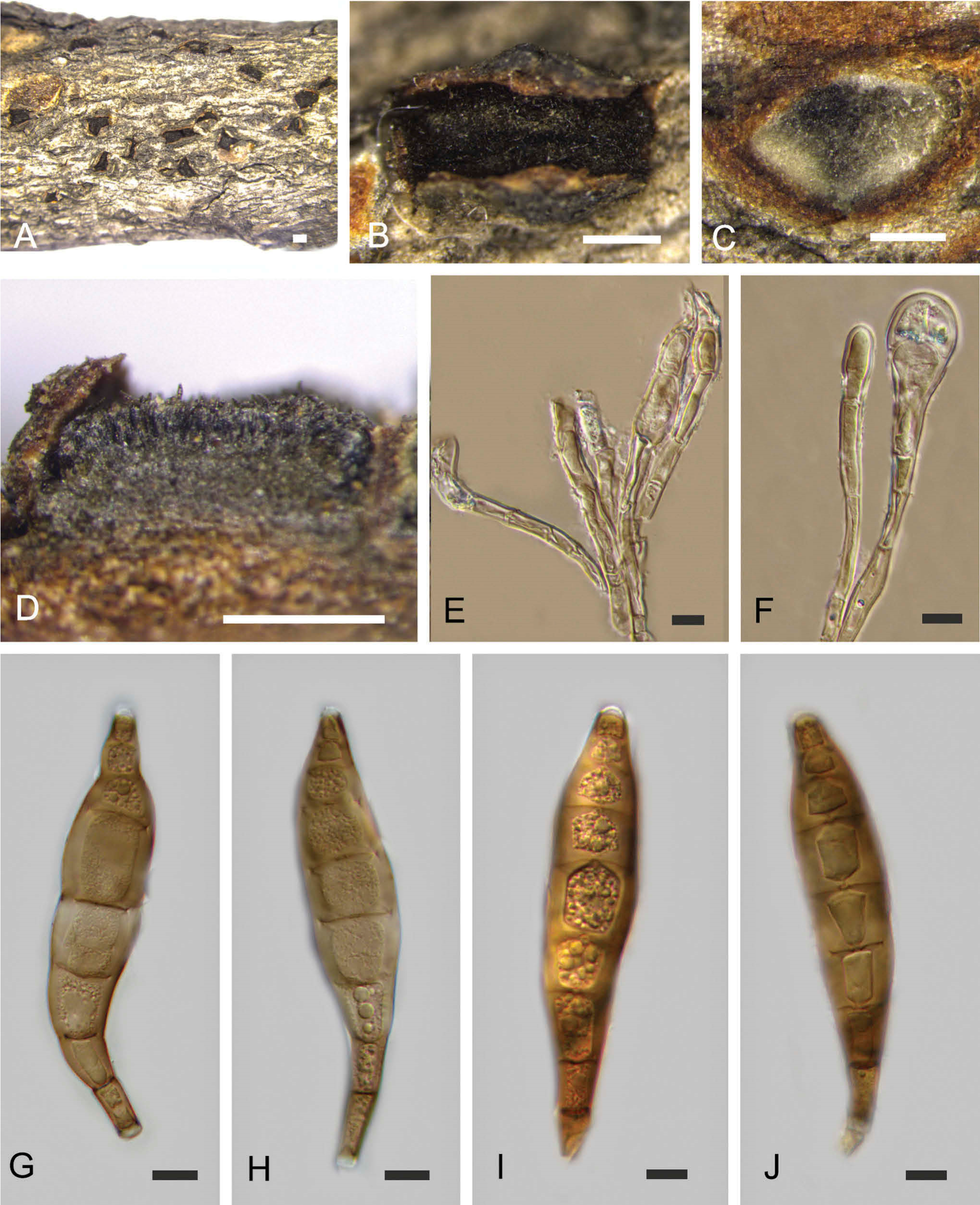
Morphology of Coryneum gigasporum from Castanea mollissima (BJFC-S1425, holotype). A, B. Conidiomata on natural substrate in surface view. C. Transverse section through conidioma. D. Longitudinal section through conidioma. E. F. Conidiophores. G–J. Conidia. Bars: A–D = 0.5 mm; E–J = 10 μm.