Cytospora xinglongensis C.M. Tian & N. Jiang, sp. nov. 2020
MycoBank No: 829517
Holotype: China, Hebei Province, Chengde City, Xinglong County, chestnut plantation, 40°24'32"N, 117°28'56"E, on branches of Castanea mollissima, 11 October 2017, N. Jiang (holotype BJFC-S1706, ex-type living culture CFCC 52458; paratype BJFC-S1707, living culture CFCC 52459).
Morphological description
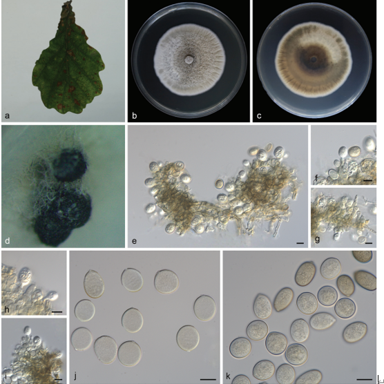
Sexual morph: not observed. Asexual morph: Pycnidial stromata immersed in bark, erumpent through the surface of bark, discoid, with a solitary undivided locule. Conceptacle black, circular surrounded stromata. Ostiole inconspicuous. Locules undivided, circular to ovoid, (480–)540–685(–755) µm diam. Conidiophores hyaline, unbranched. Peridium comprising a few layers of cells of textura angularis, with innermost layer brown, outer layer brown to dark brown. Conidiogenous cells enteroblastic polyphialidic, (4.5–)6.5–8.5(–12) × 1–1.5 µm (‒x = 7.4 × 1.3 µm). Conidia hyaline, allantoid, eguttulate, smooth, aseptate, thin-walled, (7.5–)8.5–9.5(–10.5) × 1–1.5 µm (‒x = 8.9 × 1.3 µm). Culture characters. On PDA at 25 °C in darkness. Cultures are white. The colony is flat, thin with a uniform texture, lacking aerial mycelium. Pycnidia distributed uniformly on medium surface.
Habitat: on branches of Castanea mollissima.
Distribution: Hebei Province, China.
GenBank Accession:
Notes: Cytospora xinglongensis is associated with canker disease of Castanea mollissima in China. Cytospora xinglongensis can be distinguished from its phylogenetically closely species C. thailandica by having much longer conidia (8.5–9.5 µm in C. xinglongensis vs. 3.3–4 µm in C. thailandica) (Norphanphoun et al. 2018). In addition, Cytospora xinglongensis differs from C. thailandica by ITS, ACT and RPB2 loci (16/470 in ITS, 22/245 in ACT and 52/726 in RPB2).
Reference: Jiang N, Yang Q, Fan X-L et al. (2020) Identification of six Cytospora species on Chinese chestnut in China.
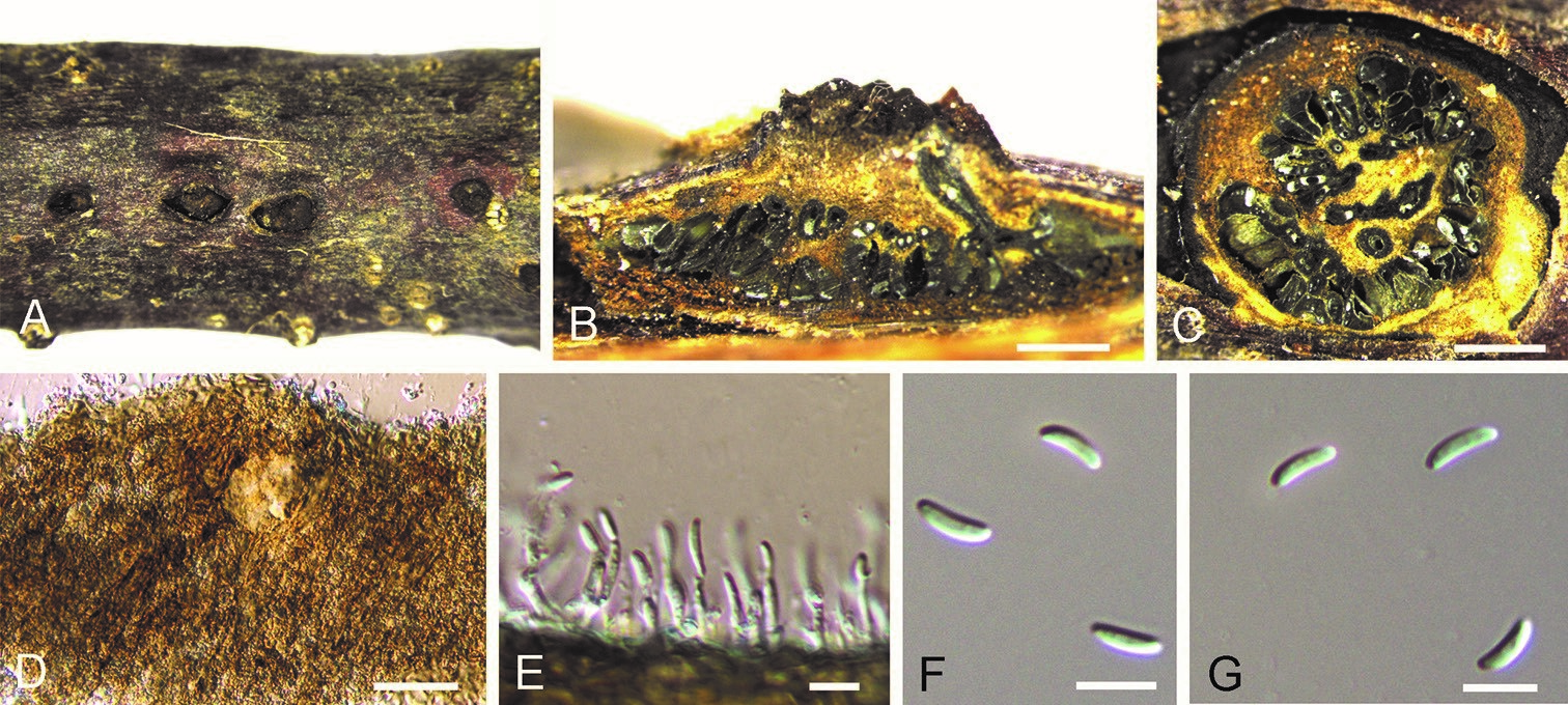
Cytospora xinglongensis on Castanea mollissima (BJFC-S1706). A Habit of conidiomata on branches B longitudinal section through conidiomata C transverse section of conidiomata D peridium E conidiogenous cells attached with conidia F, G conidia. Scale bars: 500 µm (B, C), 10 µm (E–G).