Diaporthe spinosa Y.S. Guo & G.P. Wang 2020
MycoBank MB830659
Holotype: China, Jiangsu Province, Nanjing City, on branches of P. pyrifolia cv. Cuiguan, 22 Aug. 2016, Y.S. Guo (holotype HMAS 248151, culture ex-type CGMCC 3.19602 = PCSG 383); ibid., culture PCSG 388; Zhejiang Province, Hangzhou City, on branches of P. pyrifolia cv. Cuiguan, 7 Mar. 2016, Y.S. Guo (PCSG 279); Guizhou Province, Guizhou City, on branches of P. pyrifolia cv. Yuanhuang, 8 Nov. 2017, Y.S. Guo (PCSG 491).
Morphological description
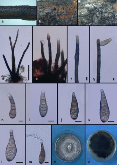
Sexual morph on fennel stems. Ascomata black, deeply embedded in fennel stems surface, 702–1404 mm diam, densely clustered in groups, multiple tapering spiny perithecial necks protruding through substrata, 1235–1864 mm long. Perithecia oval to subglobose, dark brown, 67–215 μm, ostiolate. Asci fasciculate, unitunicate, 30.5–38.5 × 6–9 μm, 8- spored, sessile, elongate to clavate. Ascospores hyaline, two-celled, often biguttulate, elliptical to fusiform, 9.5–11.5 × 3–4 μm, mean ± SD = 10.5 ± 0.6 × 3.4 ± 0.3 μm, L/W ratio = 3.1 (n = 30). Asexual morph on alfalfa stems. Pycnidial conidiomata globose, solitary, exposed on the alfalfa stems surface, dark brown to black, 124–172 μm diam. Conidiophores hyaline, smooth, 1-septate, densely aggregated, unbranched, ampulliform, 6–9 × 3–4.5 μm. Conidiogenous cells phialidic, hyaline, terminal, cylindrical, straight, 8–29 × 1.5–2.5 μm, tapered towards the apex. Alpha conidia hyaline, aseptate, fusiform to oval, acutely round at both ends, biguttulate or multi-guttulate, 5.5–8 × 2–3.5 μm, mean ± SD = 7 ± 0.6 × 2.6± 0.3 μm, L/W ratio = 2.7 (n = 50). Beta conidia hyaline, aseptate, multi-guttulate, filiform, curved, tapering towards both ends, 18.5–30.5 × 1–1.5 μm, mean ± SD = 25.1 ± 2.8 × 1.3 ± 0.1 μm, L/W ratio = 19.3 (n = 38). Gamma conidia not observed.
Culture characteristics — Colony on PDA with fluffy mycelium, panniform, aerial mycelium white, reverse umber coloured, being darker at the centre and lighter at the edge. Colony diam 62.5–67.5 mm in 3 d at 28 °C. On OA, colony with entire margin, citrine green in the centre with a white margin.
Habitat: On branches of P. pyrifolia cv. Cuiguan.
Distribution: In China.
GenBank Accession: ITS: MK626849; CAL: MK691129; HIS: MK726156; TEF: MK654811; TUB: MK691234
Notes: Diaporthe spinosa forms a well-supported, independent clade in the D. arecae species complex (Fig. 4). It contains four isolates which are separated into two branches, with the former (PSCG 383, PSCG 279) differing from the latter (PSCG 388, PSCG 491) by unique fixed alleles in three loci including ITS positions 340 (C), 342 (G), 346 (A), 347 (A), 349 (G), 380 (T), CAL positions 368 (G), and HIS positions 162 (C), 163 (A), 191 (T), 193 (C), 194 (C), 195 (T), 205 (A), 213 (C), 404 (C), 417 (T), but without obvious differences in morphology of the asexual morph. Diaporthe spinosa is most closely related to D. pescicola and D. fulvicolor, but D. spinosa and D. pescicola can be clearly differentiated from the latter by 43 different unique fixed alleles in CAL loci, and 15 different unique fixed alleles in CAL loci can also distinguish D. spinosa from D. fulvicolor. This species differs from D. pescicola in its smaller conidiomata (124–172 vs 637–881 μm), and from D. fulvicolor in its shorter alpha conidia (5.5–8 × 2–3.5 vs 7–9 × 2–3 μm).
Reference: Y.S. Guo1,2,3,4, P.W. Crous 5,6,7,8, Q. Bai 4 et al.
Diaporthe spinosa. a–d. Front and back view, respectively of colonies on PDA (a, b) and OA (c, d); e. ascomata on alfalfa stems; f–g. ascomata; h. perithecial neck; i. ascoma; j. section view of ascoma; k. asci; l. ascospores; m. conidiomata on alfalfa stems; n. conidiomata; o. section view of conidiomata; p–q. conidiophores; r. alpha conidia; s. beta conidia; t. alpha and beta conidia (a–d, m–t. isolate PSCG 383; e–l. PSCG 491). — Scale bars: e, m = 2 mm; f–g, n = 500 μm; h–j, o = 50 μm; k–l, p–t = 10 μm.