Diaporthe zaobaisu Y.S. Guo & G.P. Wang 2020
MycoBank MB830660
Holotype: China, Yunnan Province, Kunming City, on branches of P. bretschneideri cv. Zaobaisu, 17 Oct. 2014, Q. Bai (holotype HMAS 248152, culture ex-type CGMCC 3.19598 = PSCG 031); ibid., culture PSCG 032 and PSCG 033.
Morphological description
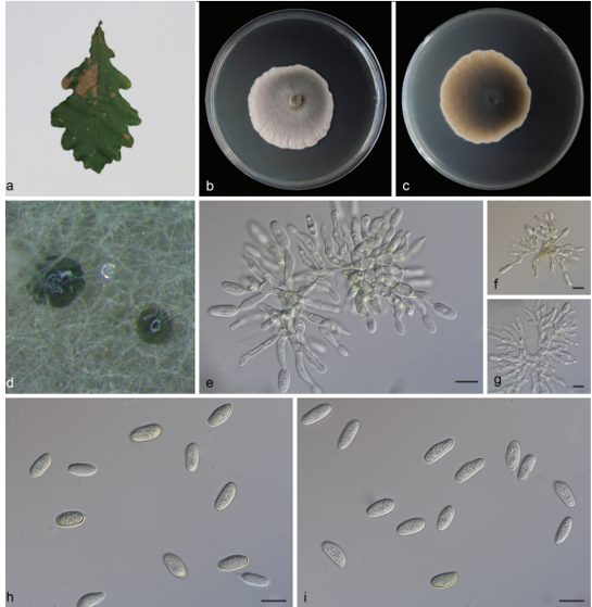
Sexual morph not observed. Asexual morph on alfalfa stems. Pycnidial conidiomata globose or irregular, solitary or aggregated, exposed on the alfalfa stems surface, dark brown to black, 235–445 μm diam. Conidiophores hyaline, smooth, 1-septate, densely aggregated, cylindrical, straight, 6–13 × 2.5–4 μm. Conidiogenous cells phialidic, hyaline, terminal, ampulliform, 8.5–12 × 2.5–3 μm, tapered towards the apex. Alpha conidia hyaline, aseptate, fusiform, biguttulate, 5.5–8.5 × 2–3 μm, mean ± SD = 6.4 ± 0.7 × 2.3 ± 0.2 μm, L/W ratio = 2.8 (n = 50). Beta conidia hyaline, aseptate, filiform, curved, tapering towards both ends, 21.5–28 × 1–1.4 μm, mean ± SD = 24.5 ± 1.5 × 1.1 ± 0.1 μm, L/W ratio = 22.3 (n = 41). Gamma conidia not observed.
Culture characteristics — Colonies on PDA flat with entire margin, colony honey in the centre with fluffy aerial mycelia and pale white margin; reverse with dull green pigment in the centre. Colony diam 40–44 mm in 3 d at 28 °C. On OA, colonies cottony, dense, greenish olivaceous in the centre; reverse dark herbage green.
Habitat: On branches of P. bretschneideri cv. Zaobaisu.
Distribution: In China.
GenBank Accession: ITS: MK626922; HIS: MK726207; TEF : MK654855 ;TUB: MK691245
Notes: The three isolates studied form a well-supported independent clade distinct from known Diaporthe species. Diaporthe zaobaisu is most closely related to D. baccae, D. rhusi- cola, D. foeniculina, D. neotheicola and D. ravennica, but differentiated from them in ITS (9 different unique fixed alleles by D. baccae, 5 by D. rhusicola, 11 by D. foeniculina, 11 by D. neo- theicola and 2 by D. ravennica) and TEF loci (21 different unique fixed alleles by D. baccae, 20 by D. rhusicola, 20 by D. foeniculina, 28 by D. neotheicola and 20 by D. ravennica). Moreover, D. zaobaisu differs from D. baccae in having shorter conidiophores (6–13 × 2.5–4 vs 20–57 × 2–3 μm) and conidiogenous cells (8.5–12 × 2.5–3 vs 9–23 × 1–2 μm) (Lombard et al. 2014). Alpha conidia are smaller than in D. foeniculina (5.5–8.5 × 2–3 vs 8.5–9 × 2–2.5 μm) and D. ravennica (5.5–8.5 × 2–3 vs 7–10.5 × 1.5–3 μm) (Udayanga et al. 2014a, Thambugala et al. 2016). Pycnidial conidiomata are smaller than in D. foeniculina (235–445 vs 400–700 μm) and D. neotheicola (235–445 vs 420–730 μm) (Santos & Phillips 2009, Udayanga et al. 2014a).
Reference: Y.S. Guo1,2,3,4, P.W. Crous 5,6,7,8, Q. Bai 4 et al.

Diaporthe zaobaisu. a–d. Front and back view, respectively of colonies on PDA (a, b) and OA (c, d); e. conidiomata on alfalfa stems; f. conidiomata; g. conidiophores; h–i. alpha conidia; j. beta conidia; k–l. alpha and beta conidia (a–h. isolate PSCG 033; i–l. PSCG 032). — Scale bars: e = 2 mm; f = 200 μm; g = 20 μm; h, j–k = 10 μm; i = 5 μm.


