Microsporomyces pseudomagnisporus Q.M. Wang, F.Y. Bai & A.H. Li sp. nov. 2020
MycoBank MB 828820
Holotype: China, Fanjingshan Mountain, Guizhou province, obtained from a leaf of an unidentified plant, Oct. 2011, Q.-M. Wang (holotype CGMCC 2.4538T preserved in a metabolically inactive state, ex-type CBS 15746 = FJS25C3).
Morphological description
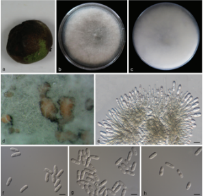
Culture characteristics: In YM broth, after 7 d at 17 °C, cells are cylindrical, 2.0–3.0 × 4.0–8.0 μm and single, budding is polar (Fig. 15I), a sediment is formed. After 1 mo at 17 °C, a part ring and sediment are present. On YM agar, after 1 mo at 17 °C, the streak culture is orange, butyrous, wrinkled and semi-glossy. The margin is entire. In Dalmau plate culture on corn meal agar, pseudohyphae are not formed. Sexual structures are not observed on YM, PDA, V8 and CM agar. Ballistoconidia are allantoid or reniform, 2.5–3.3 × 5.8–8.3 μm (Fig. 15J).
Habitat: a leaf of an unidentified plant.
Distribution: Fanjingshan Mountain, Guizhou province, China.
GenBank Accession: 18S+ITS+D1/D2 MK050384; RPB1 MK849125; RPB2 MK849351; EF1 MK849077
Notes:
Reference: A.-H. Li1,2, F.-X. Yuan1,3, M. Groenewald4 et al.