Phanerochaete metuloidea Y.L. Xu & S.H. He, sp. nov. 2020
MycoBank: MB 835449; Facesoffungi number: FoF 08035
Holotype China, Yunnan Province, Jingdong County, Ailaoshan Nature Reserve, on fallen angiosperm branch, 24 August 2015, He 2565 (BJFC 021018, holotype)
Morphological description
Fruiting body – Basidiomata annual, resupinate, widely effused, loosely adnate, easily detached from substrate, membranaceous to coriaceous, fragile after dried, up to 20 cm long, 6 cm wide. Hymenophore smooth, greyish orange [6B(3–4)], brownish orange [6C(3–4)] to light brown [6D(4–5)], turning reddish brown in KOH, uncracked or sparsely cracked with age; margin thinning out, byssoid when juvenile, becoming indistinct with age, concolorous with hymenophore surface.
Microscopic structures – Hyphal system monomitic; generative hyphae mostly simple-septate, occasionally with single or double clamp connections. Subicular hyphae colorless, slightly thickwalled, moderately branched and septate, tightly interwoven, more or less parallel to substrate, 3–7 µm in diam. Cystidia (lamprocystidia) subulate to subfusiform, colorless, thick-walled, encrusted with crystals in the upper part, with a basal simple septum, occasionally with one or two secondary septa, projecting above the hymenium, 40–80 × 5–8 µm. Basidia clavate, colorless, thin-walled, with a basal simple septum and four sterigmata, 40–70 × 5–8.5 µm; basidioles numerous, similar to basidia but smaller. Basidiospores ellipsoid, colorless, thin-walled, smooth, IKI–, CB–, (4.5–) 5–6 × 2.5–3 µm, L = 5.5 µm, W = 2.8 µm, Q = 2 (n = 30/1).
Habitat: on fallen angiosperm branch
Distribution: Southern China, mainly on Quercus.
GenBank Accession: ITS MT235682 nLSU MT248163\ MT248164 Literature This study
Notes: Phanerochaete metuloidea is characterized by the presence of thick-walled lamprocystidia and long basidia. In macro-morphology, P. metuloidea is similar to P. laevis, P. sordida and P. burdsallii, but differs in having soft texture basidiomata, obviously thick-walled cystidia, and much longer basidia. In the phylogenetic tree, two samples of P. metuloidea formed a lineage sister to P. laevis and P. burdsallii, but distant from P. sordida (Fig. 1). Phanerochaete livescens (P. Karst.) Volobuev & Spirin is similar to P. metuloidea by sharing thick-walled lamprocystidia, but differs in having ceraceous, ochraceous basidiomata, smaller basidia (16.4–27.2
× 3.9–5.0 μm) and a northern temperate distribution (Volobuev et al. 2015). Phanerochaete australis Jülich differs from P. metuloidea in having shorter but wider cystidia (38–56 × 7–10.8 μm) and shorter basidia (20–25.8 × 4.3–5 μm, Xiong & Dai 2009).
.
Reference: Xu YL1 , Cao YF1 , Nakasone KK2 et al.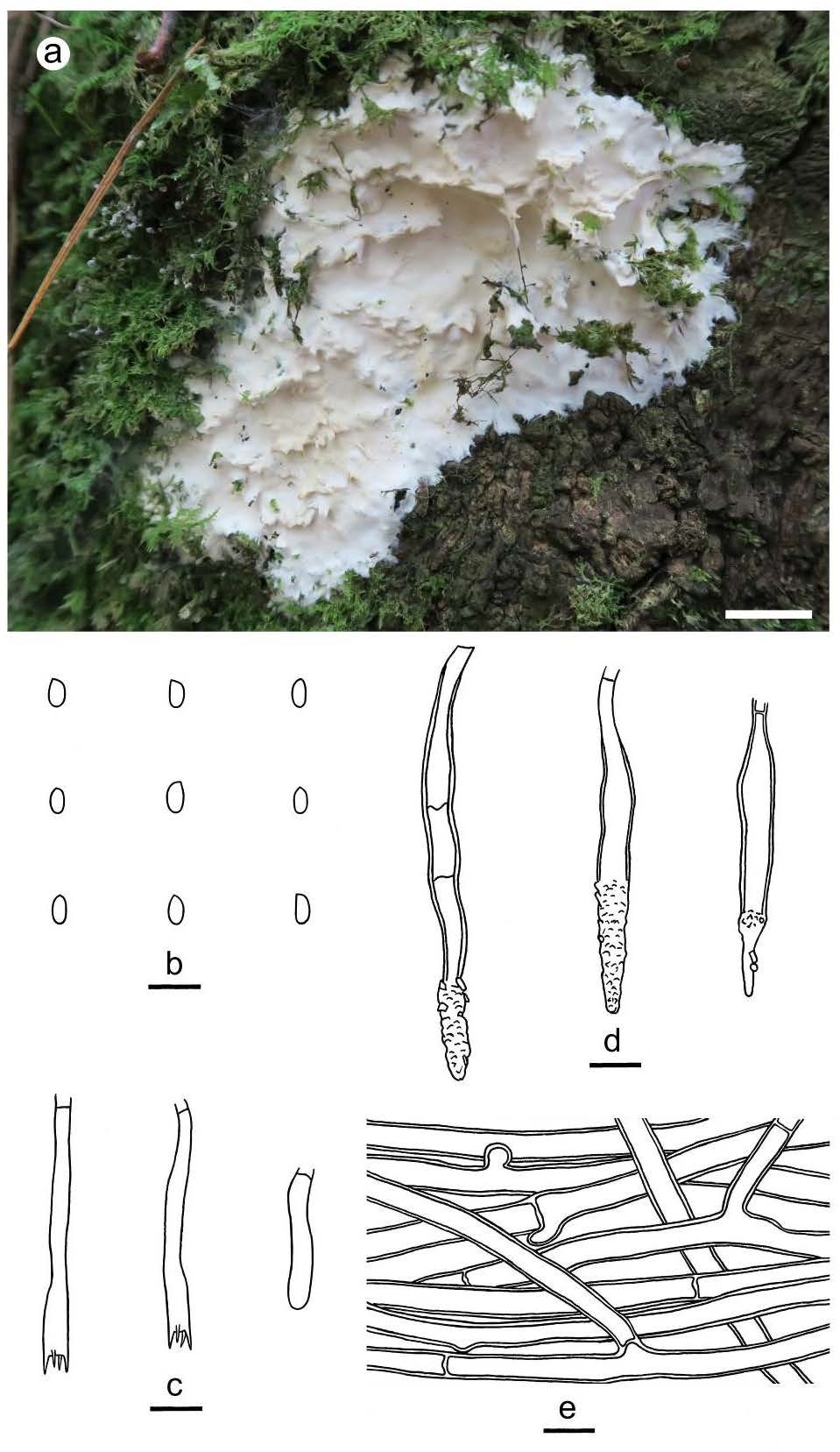
Figure 6 – Phanerochaete metuloidea (a from He 3235, b–e from the holotype He 2565). a basidiomata. b basidiospores. c basidia and a basidiole. d cystidia. e hyphae from subiculum. Scale bars: a = 1 cm, b–e = 10 µm.