Phanerochaete leptocystidiata Y.L. Xu & S.H. He, sp. nov.2020
MycoBank: MB 835448; Facesoffungi number: FoF 08034
Holotype :China, Guangdong Province, Renhua County, Danxiashan Nature Reserve, on fallen angiosperm trunk, 4 June 2019, He 5853 (BJFC 030728, holotype).
Morphological description
Fruiting body – Basidiomata annual, resupinate, effused, loosely adnate, easily detached from substrate, pellicular to membranaceous, up to 10 µm long, 8 cm wide. Hymenophore smooth to tuberculate, white (6A1) when fresh, becoming smooth, pale orange (6A3), orange grey (6B2) to greyish orange [6B(3–5)] when dry, slightly darkening in KOH, sparsely cracked with age; margin thinning out, fibrillose, concolorous with hymenophore surface; hyphal cords greyish orange [6B(4–5)] to orange [6B(7–8)], turning reddish brown in KOH.
Microscopic structures – Hyphal system monomitic; generative hyphae mostly simple-septate, occasionally with single or double clamp connections. Subicular hyphae colorless, slightly thickwalled, encrusted with crystals, rarely branched and septate, loosely interwoven, more or less parallel to substrate, 3–7 µm in diam. Cystidia subulate, tapered toward apex, colorless, thin-walled, smooth or rarely encrusted at apex, projecting above the hymenium, with a basal simple septum, 30–70 × 4–6 µm. Basidia clavate, colorless, thin-walled, with a basal simple septum and four sterigmata, 24–30 × 4.5–6 µm; basidioles numerous, similar to basidia but smaller. Basidiospores ellipsoid to subcylindrical, colorless, thin-walled, smooth, IKI–, CB–, 5–6 (–6.5) × 2.5–3 µm, L = 5.5 µm, W = 2.7 µm, Q = 2.1 (n = 30/1).
Habitat: on fallen angiosperm trunk
Distribution: Guangdong, Guizhou and Jiangxi Provinces, southern subtropical China.
GenBank Accession: ITS MT235685\ MT235684 nLSU MT248168\ MT248167 Literature This study\ This study
Notes: Phanerochaete leptocystidiata is characterized by the white to greyish orange pellicular basidiomata with hyphal cords and leptocystidia. In the phylogenetic tree, P. leptocystidiata, P. sinensis and P. burtii (Romell ex Burt) Parmasto formed a strongly supported clade (Fig. 1). Morphologically, the three species are very similar to each other by sharing pellicular basidiomata with hyphal cords and leptocystidia. Phanerochaete sinensis differs from P. leptocystidiata by having slightly shorter cystidia (35–50 µm), slightly smaller basidiospores (4–5 × 2–2.5 µm) and a temperate distribution. Phanerochaete burtii differs from P. leptocystidiata by slightly shorter cystidia (25–55 µm), slightly narrower basidiospores (2–2.5 µm) and a distribution in U.S.A., Jamaica, Argentina, Brazil and Australia (Burdsall 1985, Hjortstam 2000). The P. burtii group is also morphologically similar to and phylogenetically close to P. sanguinea (Fr.) Pouzar group and P. carnosa (Burt) Parmasto. However, species of P. sanguinea group have orange to red basidiomata that usually stain the substrate with same color, whilst P. carnosa has an ochraceous hymenophore that turns dark green in KOH (Burdsall 1985, Xiong & Dai 2009, Floudas & Hibbett 2015).
Reference: Xu YL1 , Cao YF1 , Nakasone KK2 et al.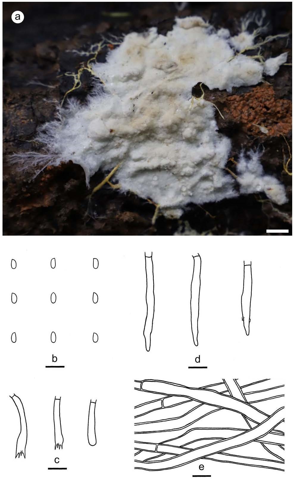
Figure 5 – Phanerochaete leptocystidiata (from the holotype He 5853). a basidiomata. b basidiospores. c basidia and a basidiole. d cystidia. e hyphae from subiculum. Scale bars: a = 1 cm, b–e =10 µm.