Cytospora kuanchengensis C.M. Tian & N. Jiang 2020
MycoBank No: 829514
Holotype: . China, Hebei Province, Chengde City, Kuancheng County, chestnut plantation, 40°38'37"N, 118°27'54"E, on branches of Castanea mollissima, 13 October 2017, N. Jiang (holotype BJFC-S1695, ex-type living culture CFCC 52464; paratype BJFC-S1696, living culture CFCC 52465).
Morphological description
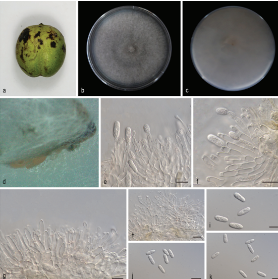
Sexual morph: not observed. Asexual morph: Pycnidial stromata ostiolated, immersed in bark, scattered, erumpent through the surface of bark, discoid, with multiple locules. Conceptacle black, circular surrounded stromata. Ectostromatic disc black, circular to ovoid, (350–)455–540(–575) µm diam., with 1–7 ostiole per disc. Ostioles black, at the same level as the disc, (40–)60– 85(–115) µm diam. Locule numerous, arranged circularly or elliptically with independent walls, (285–)355–520(–605) µm diam. Peridium comprising few layers of cells of textura angularis, with innermost layer brown, outer layer brown to dark brown. Conidiophores hyaline, unbranched, thin walled, filamentous. Conidiogenous cells enteroblastic polyphialidic, (6.5–)8.5–11(–15) × 1–1.5 µm (x‒ = 9.8 × 1.3 µm). Conidia hyaline, allantoid, smooth, aseptate, thin-walled, (5.5–)6–7.5(–8) × 1–2 µm (x‒ = 6.9 × 1.6 µm).Culture characters. On PDA at 25 °C in darkness. Cultures are initially white, producing pale brown pigment after 10 d. The colony is flat, felt-like, with concentric circular texture. Pycnidia distributed irregularly on medium surface.
Habitat: on branches of Castanea mollissima.
Distribution: China.
GenBank Accession: ITS:MK432616;LSU: MK429886;ACT: MK442940;RPB2: MK578076
Notes: Cytospora kuanchengensis is associated with canker disease of Castanea mollissima in China. Cytospora kuanchengensis differs from its phylogenetically closely species, C. pruinosa, by ITS and ACT loci (7/470 in ITS and 21/245 in ACT). Morphologically, C. kuanchengensis has slightly larger conidia than C. pruinose (5.5–8 × 1–2 µm in Cytospora kuanchengensis vs. 5–7.5 × 1–1.5 µm in C. pruinosa) (Fan et al. 2020).
Reference: Jiang N, Yang Q, Fan X-L et al. (2020) Identification of six Cytospora species on Chinese chestnut in China. MycoKeys 62: 1–25. https://doi.org/10.3897/mycokeys.62.47425
Cytospora kuanchengensis on Castanea mollissima (BJFC-S1695). A, B Habit of conidiomata on branches C longitudinal section through conidiomata D transverse section of conidiomata E peridium F, G conidiogenous cells attached with conidia H conidia. Scale bars: 500 µm (B–D), 10 µm (E–G), 5 µm (H).