Cytospora ceratospermopsis C.M. Tian & X.L. Fan, Persoonia. 2020
MycoBank
Holotype: China, Hebei Province, Chengde City, Xinglong County, chestnut plantation, 40°24'32"N, 117°27'56"E, on branches of Castanea mollissima, 11 October 2017, N. Jiang (BJFC-S1699, living culture CFCC 52471 from the ascospore; BJFC-S1700, living culture CFCC 52472 from the conidium).
Morphological description
Sexual morph: Ascostromata immersed in the bark, erumpent through the surface of bark, scattered, (350–)550–900(–1300) µm diam., with 15–40 perithecia arranged circularly or irregularly. Conceptacle absent. Ectostromatic disc black, usually surrounded by tightly ostiolar necks, circular to ovoid, (180–)240–410(–450) µm diam. Ostioles black, at the same level as the disc or slightly above, concentrated, dark brown to black, arranged circularly in a disc, (55–)60–85(–110) µm diam. Perithecia dark brown, flask-shaped to spherical, arranged circularly or irregularly, (255–)280–350(–420) µm diam. Asci clavate to elongate obovoid, 8-spored, (20.5–)27–35.5(–43) × (4–)4.5–6.5(– 8) µm (x‒= 31.2 × 5.6 µm). Ascospores biseriate, elongate-allantoid, thin-walled, hyaline, aseptate, (5.8–)7.5–9.2(–11.5) × (3–)3.2–4.8(–5.5) µm (x‒ = 8.6 × 4.1 µm). Asexual morph: Pycnidial stromata ostiolated, immersed in bark, scattered, erumpent through the surface of bark, discoid to conical, with multiple locules. Conceptacle absent. Ectostromatic disc light brown to grey, circular to ovoid, (230–)280–360(–480) µm diam., with one ostiole per disc. Ostiole in the centre of the disc, dark grey to black, conspicuous, at the same level as the disc, (60–)75–110(–135) µm diam. Locule numerous, arranged circularly or elliptically with independent walls, (300–)350–600(–950) µm diam. Peridium comprising few layers of cells of textura angularis, with innermost layer brown, outer layer brown to dark brown. Conidiophores hyaline, branched or not, thin walled, filamentous. Conidiogenous cells enteroblastic polyphialidic, (6.5–)8.5–15.5(– 18) × 1.5–2.5 µm (x‒ = 12.2 × 1.9 µm). Conidia hyaline, allantoid, smooth, aseptate, thin-wall, (4.5–)5–6.5(–7) × 1–1.5 µm (x‒ = 5.9 × 1.3 µm).Culture characters. On PDA at 25 °C in darkness. Cultures are initially white, becoming olivaceous buff in centre after 7 d and finally olivaceous at 30 d. The colony is flat, thin with a felt and tight texture in centre. Pycnidia distributed irregularly on medium surface.
Habitat: on branches of Castanea mollissima
Distribution: China.
GenBank Accession: ITS: MK432629; LSU: MK429899;ACT: MK442953;RPB2: MK578087
Notes: Fresh specimens with both sexual and asexual morphs were collected from cankered branches of Castanea mollissima and two isolates were obtained from the ascospore and conidium, respectively. Phylogenically, the two isolates were close to Cytospora ceratospermopsis represented by CFCC 89626 and CFCC 89627 (Fig. 2). We compared their sequences and found no differences in LSU and RPB2, but 2 bp differences in ITS and 3 bp differences in ACT. Fan et al. (2020) reported the asexual morph of Cytospora ceratospermopsis from Juglans regia in China with conidial size in 4.5–6 × 1–1.5 µm, which is exactly matched with the asexual characters observed in the present study. Hence, we described the asexual morph of Cytospora ceratospermopsis in its sexual morph for the first time and reported a new host, Castanea mollissima.
Reference: Jiang N, Yang Q, Fan X-L et al.(2020) Identification of six Cytospora species on Chinese chestnut in China. MycoKeys 62: 1–25. https://doi.org/10.3897/mycokeys.62.47425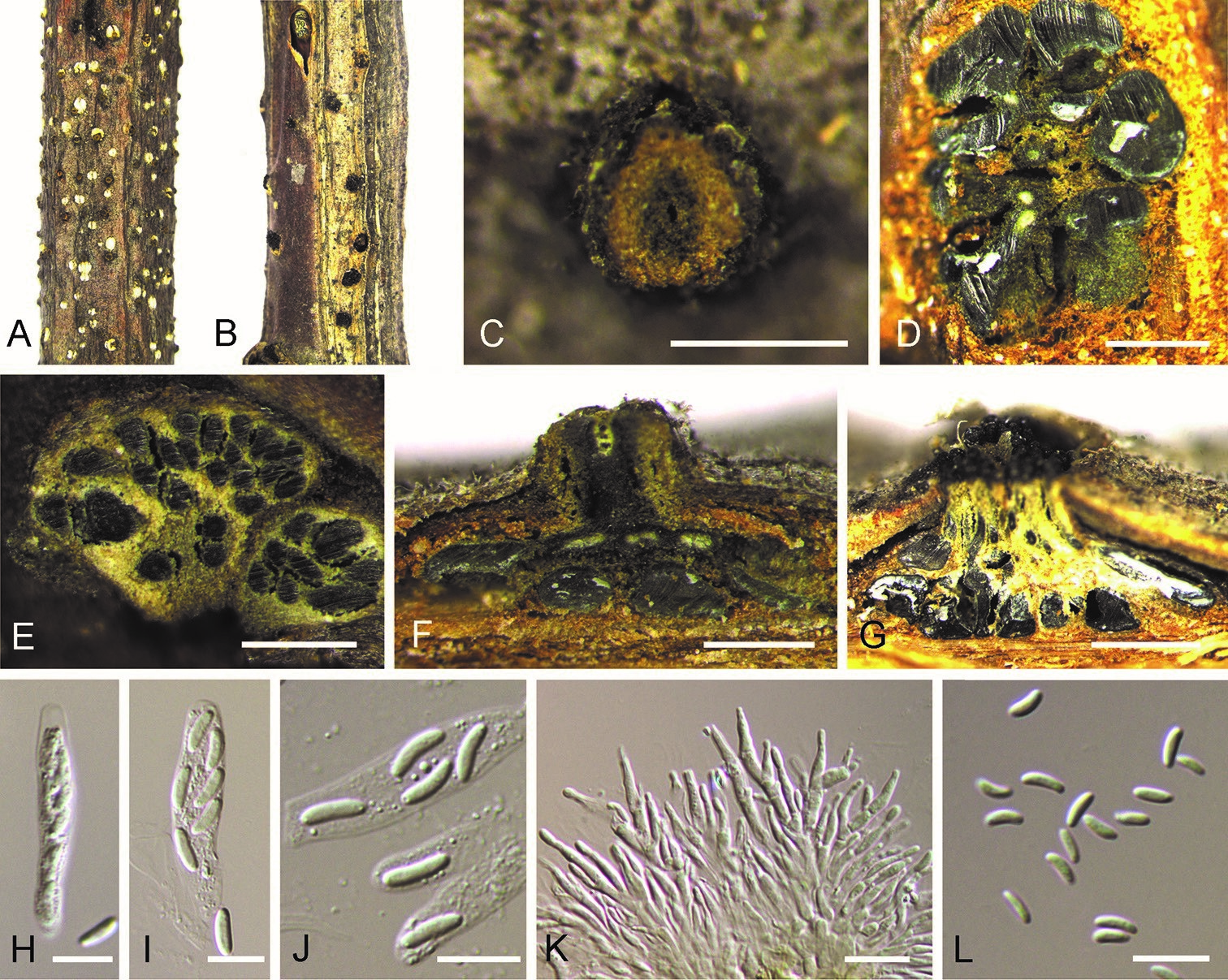
Cytospora ceratospermopsis on Castanea mollissima (BJFC-S1699, BJFC-S1700). A, C Habit of conidiomata on branches B habit of ascomata on branches D transverse section of conidiomata E transverse section of ascomata F longitudinal section through conidiomata G longitudinal section through ascomata H, I asci J ascospores K conidiogenous cells with attached conidia L conidia. Scale bars: 500 µm (C–G), 10 µm (H–L).