Torula camporesii Phookamsak, E.F. Yang & K.D. Hyde 2020
Index Fungorum number: IF557230; Facesoffungi number: FoF 07198
Holotype: CHINA, Yunnan Province, Xishaungbanna, Mengla Country, Bubeng Field Station, Xishuangbanna Station for Tropical Rain Forest Ecosystem Studies (101° 35′ 4″ E, 21° 36′ 4″ N, ± 683 msl.), on decaying herbaceous plant, 7 May 2018, R. Phookamsak, XB001 (MFLU 20-0070, holotype), ex-type living culture, KUMCC 19-0112.
Morphological description
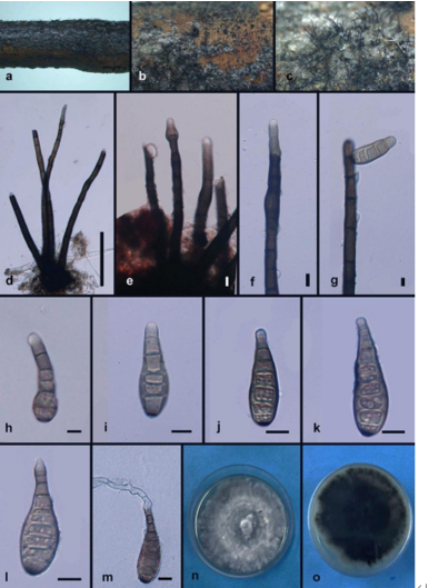
Sexual morph Undetermined. Asexual morph Colonies visible as black dense, velvety, powdery on host. Mycelium 1.5–3.5 μm in wide, partly immersed to superficial on the substrate, composed of septate, branched, light brown hyphae. Conidiophores 9–18.5 × 3–5 μm ( x̄ = 14 × 4 μm, n = 50), macronematous to semi- macronematous, mononematous, erect, straight, or slightly flexuous, without apical branches, light brown to brown, ellipsoid to subcylindrical, 0–1-septate, smoothwalled, with 1–3, globlose to subglobse cells, arising from prostrate hyphae. Conidiogenous cells 5.5–8.5 × 5–8 μm ( x̄ = 7 × 6.5 μm, n = 30), mono- to polyblastic, integrated, terminal or intercalary, cupulate to globose or subglobose, brown to dark brown, smooth to verrucose thick-walled. Conidia (34–)40–70(–77) × 6–8(–10) μm ( x̄ = 53.9 × 6.9 μm, n = 50), catenated, composed of moniliform cells, acrogenous, phragmosporous to scolecosporous, brown to dark brown, verruculose, wider cells in the middle, often smaller at apex, 4–20-septate, constricted at the septa, chiefly subcylindrical, produced branched chain, globlose to subglobose of each cell, basal cell slightly truncate, with a darkened scar at the base. Conidial secession schizolytic.Culture characteristics: Colones on PDA, reaching around 40 mm diam., after 2 weeks at room temperature (20–25 °C), colonies medium dense, irregular, slightly effuse, rough, with standing tufts at the centre, umbonate edge, colonies from above, white to cream at the margin, pinkish white to white-brown at the centre, gradually turn to brown when mature, from below white to pink-white at the margin, irregular radiating with pink-brown at the middle, reddish brown to pinkish brown at the centre; not producing pigment in PDA.
Habitat: herbaceous litter.
Distribution:China.
GenBank Accession: I TS:M N 5 0 7 4 0 0;LSU:MN507402;SSU:MN507401;RPB2:MN507404;TEF1-α:MN507403.
Notes: Multi-gene phylogenetic analyses of a combined LSU, SSU, TEF1-α, RPB2 and ITS sequence dataset indicated that Torula camporesii forms a sister lineage with T. goaensis Pratibha & Prabhugaonkar in Torula with high support (100% ML, 1.00 BYPP; Fig. 80). Torula camporesii resembles T. goaensis in having phragmosporous to scolecosporous, brown, verruculose, 4–20-septate conidia. However, the species can be distinguished from T. goaensis in having shorter conidial chains (T. camporesii, (34–)40–70(–77) × 6–8(–10) versus 42–105 × 6–8.5 μm, T. goaensis) (Pratibha and Prabhugaonkar2017). Torula camporesii has conidia that are brown to dark brown, long subcylindrical to ellipsoidal in chains, wider cells in the middle, often smaller towards the end cell, basal cell slightly truncate, with a darkened scar at the base. Torula goaensis has light brown to brown conidia, obtuse at the basal cell, and without a darkened scar at the base (Pratibha and Prabhugaonkar2017). In NCBI BLASTn search based on ITS sequences, the closest match of T. camporesii is T. goaensis (MTCC 12620; GenBank no. NR_159045) with 97.14% similarity. A comparison of ITS nucleotide bases indicate that T. camporesii differs from T. goaensis in 15/522 bp (2.87%). Therefore, T. camporesii is introduced as a novel species based on the justification guildelines of Jeewon and Hyde (2016).
Reference: Kevin D. Hyde1,5,8,22 · Yang Dong2,3 · Rungtiwa Phookamsak1,5,6,7 et al.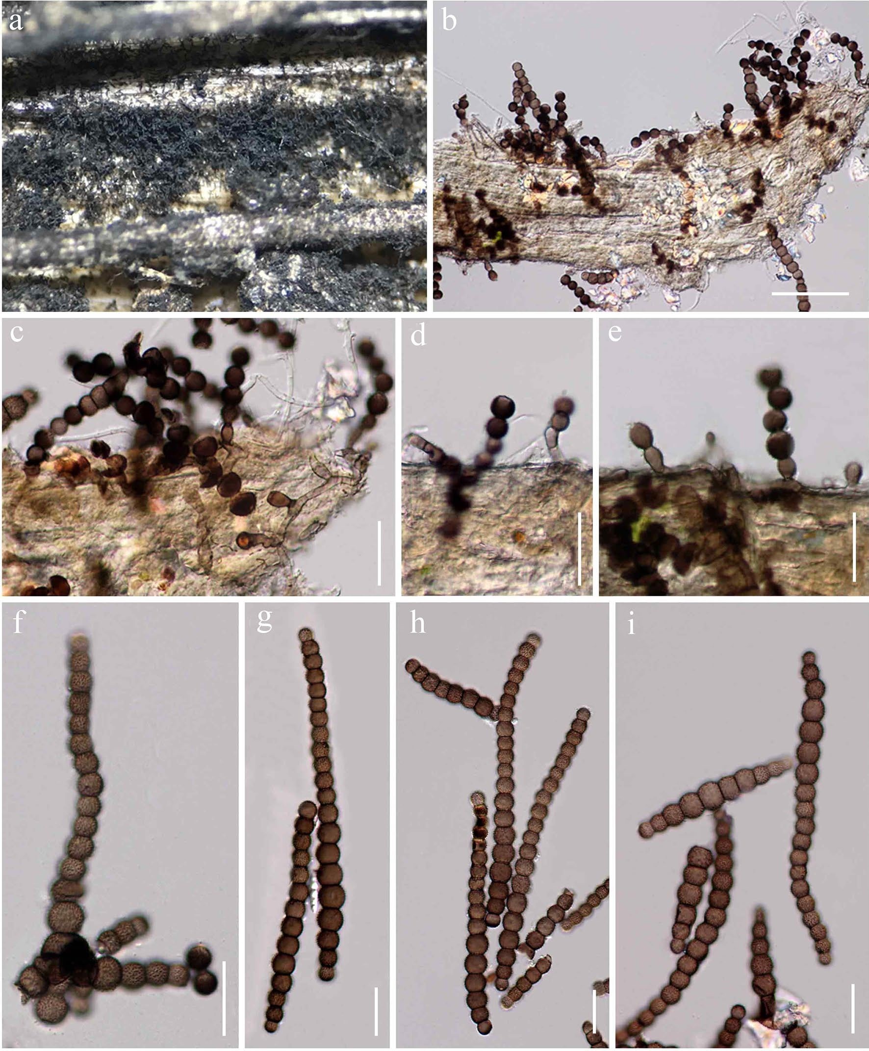
Torula camporesii (MFLU 20-0070, holotype). a Appearance of colonies on host substrate. b, c Colonies on host substrate showing partly immersed mycelium and its erect conidiophores attached with conidia. d, e Conidiophores and conidiogenous cells. f–i Conidia. Scale bars: b=50 μm, c–i=20 μm