Trichomerium cicatricatum L. Su, W. Sun and M.C. Xiang, sp. nov. 2020
MycoBank MB 810890
Holotype: China: Jiangxi Province: Jiujiang City, Guling Town, Lushan Mountain, from sandstone, climate with abundant rainfall and sunshine, 29◦360 N, 115◦590 E, 455 m a.s.l., 19 May 2013, Lei Su, (HMAS245352 (dried culture)–holotype and CGMCC 3.17307–ex-type culture).
Morphological description
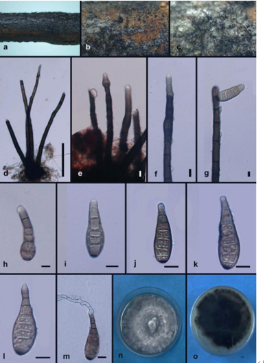
Hyphae cylindrical, branched, septate, constricted at the septa, 2–6-µm-wide, brown to dark brown, oil drops abundant in young hyphae, enteroblastic proliferation common at the ends of moniliform hyphae, terminal cells frequently larger than the subterminal cells, 14.1–16.9-µm-long,4.9–7.0-µm-wide (x = 15.2 × 6.5 µm, n = 5), often proliferating with umbonate apex due to budding (Figure 13D–F). Endoconidia rarely present, unicellular, globose, sub-globose to ellipsoidal, 6.3–15.2 (x = 12.5 µm, n = 5)-µm-diam., light brown, usually 1-endoconidia present in individual swollen intercalary or terminal cells (Figure 13H–J, arrow). Multicellular bodies brown to dark brown, 6.9–14.9 × 10.2–16.5 µm (x = 10.5 × 13.6 µm, n = 10) (Figure 13G). Conidia Tripospermum-like, pale brown to dark brown, consisting of a subcylindrical basal cell, 1–3-septate, 20.9–33.6 × 3.2–7.5 (x = 27.6 × 5.6 µm, n = 5), with a truncate hilum, giving rise to three to four lateral arms from a central cell, arms one to two septate, subcylindrical with obtusely rounded ends, 18.5–27.8 × 2.2–7.5 (x = 22.7 × 4.9 µm, n = 5) (Figure 13C).Culture characters: Colonies on MEA growing slowly, attaining 13-mm-diam. after 20 weeks at 25 ◦C, compact, slightly raised at the center, pale black, with scarce, short, velvety aerial mycelia; margin irregular; black in reverse (Figure 13A,B). Minimum 4 ◦C, optimum at 20–25 ◦C, and maximum 28 ◦C.
Habitat: from sandstone
Distribution: China
GenBank Accession:
Notes: Phylogenetic analyses and high bootstrap support values indicated that T. cicatricatum is closely related to T. dioscoreae (MLBP/BIPP = 91%/1.00) (Figure 2). However, T. dioscoreae causes leaf spots with faster growing mycelium and smaller conidia (10–20 × 3–5 µm) [66] compared toT. cicatricatum.
Reference: Wei Sun , Lei Su , Shun Yang et al.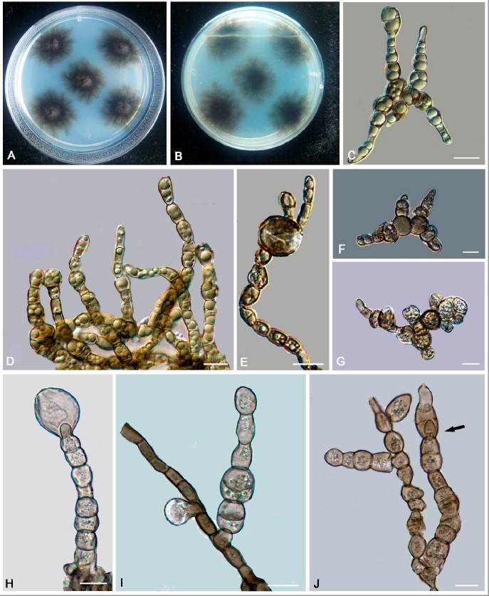
Trichomerium cicatricatum (CGMCC 3.17307). (A,B) Colony forward and reverse after
20 weeks on MEA. (C) Conidia with 4 arms. (D–F) Moniliform hyphae with proliferation at the apex. (G) Solitary, enlarged, darkly pigmented multicellular body. (H–J) Endoconidia develops within intercalary and terminal cells (arrow). Scale bars: (C–J) = 10 µm.