Lithohypha catenulata L. Su, W. Sun and M.C. Xiang, sp. nov.2020
MycoBank MB 810886
Holotype: China: Beijing: the west campus of China Agricultural University, 40◦000 N, 116◦120 E, 108 m a.s.l., from ornamental limestone, 12 April 2011, Lei Su, (HMAS264536 (dried culture)–holotype and CGMCC 3.14885–ex-type culture).
Morphological description
Hyphae moniliform, branched, smooth, pale brown to brown, diffusely guttulate, consisting of spherical to subspherical cells with enteroblastic proliferation (Figure 12C,D). Swollen cells globose to sub-globose, frequently intercalary, terminal or lateral, 6.1–10.2 µm (x = 7.5 µm, n = 20) in young cultures, pale brown, gradually turning dark brown, forming dark brown fragmented crusts on the colony surface (Figure 12E). Multicellular bodies global to ellipsoidal, brown to dark brown,
with transverse and longitudinal septa, widening up to 21.4 µm (x = 16.8 µm, n = 10) (Figure 12F–I).Culture characteristics: Colonies on MEA growing slowly, attaining 18-mm-diam. after 20 weeks at 25 ◦C, blackish brown to black, velvety, with sharp and irregularly lobed margin; black in reverse (Figure 12A,B). Minimum 4 ◦C, optimum at 20–25 ◦C, and maximum 31 ◦C.
Habitat: from ornamental limestone
Distribution: China
GenBank Accession:
Notes: Morphologically, L. catenulate share some similar characters with L. guttulata in having multicellular bodies. However, L. catenulate has transverse and longitudinal septa multicellular bodies up to 21.4 µm wide.
Reference: Wei Sun , Lei Su , Shun Yang et al.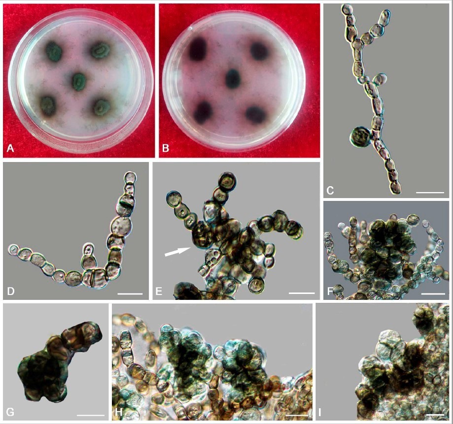
Lithohypha catenulata (CGMCC 3.14885). (A,B) Colony surface and reverse after 20 weeks on MEA. (C,D) Catenated, moniliform hyphal cells proliferating often at the apex. (E) Swollen cells with dark brown fragmented crusts on their surface (arrows). (F–I) Solitary, enlarged, dark pigmented
multicellular bodies with transverse and longitudinal septa. Scale bars: (C–I) = 10 µm.